
Acute phase protein university answer model
Reading File
Acute phase proteins (APPs) are plasma proteins whose concentrations change significantly (by at least 25%) in response to inflammation or tissue injury. Now searching the medical library for comprehensive content.
Searching sources...
Finding Sources
Finding Sources
Reading File
Reading File
Reading File
Reading File
Reading File
Reading File
Reading File
Finding Sources
Reading File
Reading File
Loading Image
Finding Sources
Reading File
Reading File
I now have comprehensive content from multiple authoritative textbooks. Let me compile the complete university-level answer.
Acute Phase Proteins (APPs) - Comprehensive University Answer
Definition
An acute phase protein is any plasma protein whose concentration changes from its baseline value by at least 25% during an inflammatory response. Proteins that increase are termed positive acute phase reactants; those that decrease are negative acute phase reactants. - Firestein & Kelley's Textbook of Rheumatology, p. 1098
The Acute Phase Response
Within minutes of tissue injury, activation of the innate immune system induces cytokine production that results in a multisystem acute phase response involving the:
- Liver (primary site of APP synthesis)
- Vascular system
- Bone marrow
- Central nervous system
This response represents a reprioritisation of hepatic protein metabolism - the liver shifts away from constitutive export proteins (albumin, transferrin) toward defence and repair proteins. - Bailey & Love's Short Practice of Surgery, 28th ed.
Induction of liver acute phase genes is mediated by the transcription factor STAT3 (Signal Transducer and Activator of Transcription 3).
Cytokine Regulation
The key cytokines driving APP synthesis in the liver are:
| Cytokine | Role |
|---|---|
| IL-6 | Primary hepatocyte-stimulating factor; signals via JAK-STAT3, C/EBP, and NF-kB pathways |
| IL-1 | Synergises with IL-6 in initial stages; triggers further IL-6 production |
| TNF-alpha | Synergises with IL-6; also drives fever, catabolism, and vascular permeability |
IL-6 is produced by activated monocytes, macrophages, neutrophils, natural killer cells, and endothelial cells. Via trans-signalling (binding soluble IL-6R and ligation of gp130), IL-6 can amplify its effects both locally and systemically. - Firestein & Kelley's Textbook of Rheumatology, p. 1098
Classification by Magnitude of Response
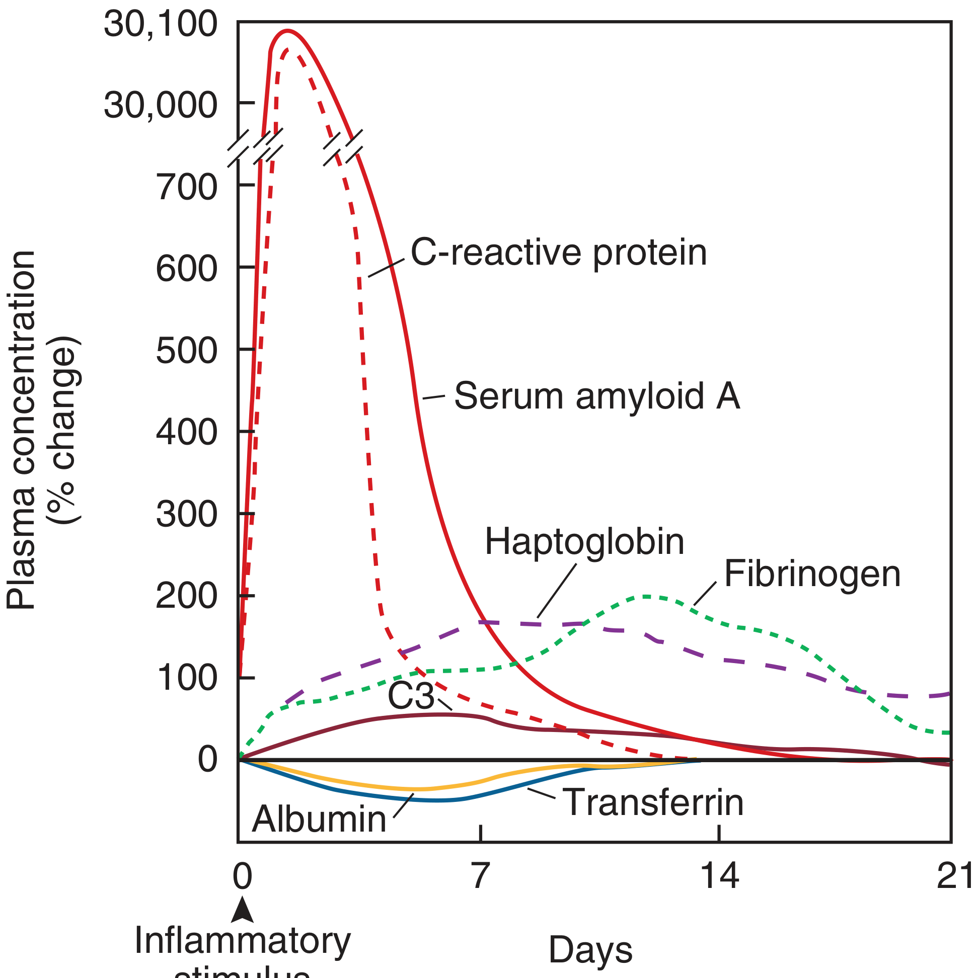
Typical plasma APP changes after a moderate inflammatory stimulus (Firestein & Kelley's Textbook of Rheumatology)
1. Major Positive APPs - Increase >1000-fold (within 2-3 days)
- C-reactive protein (CRP)
- Serum Amyloid A (SAA)
2. Moderate Positive APPs - Increase 2-4 fold (slower rise)
- Fibrinogen
- Haptoglobin
- alpha1-acid glycoprotein (orosomucoid)
- alpha1-protease inhibitor (alpha1-antitrypsin)
- alpha1-antichymotrypsin
3. Minor Positive APPs - Increase ~50-100%
- Complement C3
- Complement C9, Factor B
- Ceruloplasmin
- Ferritin
- Mannose-binding lectin (MBL)
- Fibronectin
4. Negative APPs - Decrease during inflammation
- Albumin
- Transferrin
- Pre-albumin (transthyretin)
- Apolipoprotein A-I
- Antithrombin III
- Protein S
Individual Proteins in Detail
C-Reactive Protein (CRP)
- Structure: 115 kDa pentamer of five 23 kDa subunits (non-covalently linked). Pentameric form.
- Synthesis: Liver hepatocytes. Trace baseline concentration in all humans (~0.3 mg/dL).
- Kinetics: Rises within 6-8 hours of stimulus, peaks at 2-3 days, half-life of ~19 hours. Falls rapidly once stimulus is removed.
- History: Identified in 1930 when it was found to bind the "C" polysaccharide of Streptococcus pneumoniae cell wall.
- Ligands: Binds phosphocholine, phospholipids, fibronectin, chromatin, histones (exposed on apoptotic cells and sites of tissue damage).
- Functions:
- Opsonisation: Tags pathogens and apoptotic debris for phagocytosis
- Complement activation: Activates the classical pathway
- Immune bridging: Binds Fc-gamma receptors on leukocytes
- Induction of cytokines: Promotes IL-6 receptor shedding and tissue factors
- Anti-inflammatory role: Promotes non-inflammatory clearance of apoptotic cells; prevents neutrophil adhesion to endothelium
CRP Reference Ranges and Clinical Significance:
| Level | Significance |
|---|---|
| <0.3 mg/dL | Normal (healthy adult) |
| 0.3-1 mg/dL | Minor elevation (use high-sensitivity CRP) |
| 1-10 mg/dL | Moderate elevation (significant inflammation) |
| >10 mg/dL | Marked elevation |
| >15 mg/dL | Strongly suggests bacterial infection |
| >50 mg/dL | Infection present in ~88% of cases |
Conditions with CRP elevation:
- Normal/minor (<1 mg/dL): vigorous exercise, common cold, pregnancy, gingivitis, depression, obesity, insulin resistance
- Moderate (1-10 mg/dL): myocardial infarction, malignancy, most autoimmune diseases
- Marked (>10 mg/dL): bacterial infections, severe RA flares, vasculitis
Important exceptions: CRP elevation is typically absent or modest in active SLE despite significant clinical activity - the reason remains unexplained.
- Firestein & Kelley's Textbook of Rheumatology, p. 1099-1100
Serum Amyloid A (SAA)
- Type: Apolipoprotein; also classified as an acute phase protein.
- Rise: Up to 1000-fold (same order as CRP). Associates with HDL during the acute phase response.
- Clinical use: Limited by lack of standardised assays. Can be used synergistically with hsCRP.
- Significance: Chronic elevation leads to SAA deposition as AA amyloid - the mechanism underlying secondary (reactive) amyloidosis in conditions like RA, FMF, and chronic infections.
Haptoglobin
- Function: Binds free haemoglobin to prevent oxidative damage and iron loss.
- Rise: Moderate (2-4 fold).
- Note: Low haptoglobin is a marker of intravascular haemolysis (haemoglobin binds and depletes it).
Fibrinogen
- Function: Coagulation; polymerises to fibrin clot; tissue repair.
- Rise: Moderate (2-4 fold), slower peak than CRP.
- Clinical relevance: Elevated fibrinogen contributes to increased ESR (by causing RBC rouleaux formation). Also a cardiovascular risk factor.
Ceruloplasmin
- Function: Copper transport; superoxide radical scavenger (antioxidant); ferroxidase activity.
- Rise: Minor (~50%).
- Note: Plasma copper rises during acute phase response because of increased ceruloplasmin, even though zinc and iron fall.
alpha1-Antitrypsin (alpha1-Protease Inhibitor)
- Function: Antiprotease - inhibits neutrophil elastase, collagenase, and bacterial proteases, limiting tissue destruction.
- Rise: Moderate.
alpha1-Acid Glycoprotein (Orosomucoid)
- Function: Transport protein (binds basic drugs); platelet inhibitor; immunomodulatory.
- Rise: Moderate to major.
Mannose-Binding Lectin (MBL)
- Function: Pattern recognition; fixes complement via lectin pathway; opsonisation.
- Rise: Moderate. Part of the collectin family.
Ferritin
- Function: Iron storage protein; elevated ferritin sequesters iron from microbes (bacteriostatic effect).
- Regulation: Induced by IL-1, IL-6, IL-18, TNF via NF-kB pathway.
- Hyperferritinemic syndromes: Extremely high ferritin (>10,000 ng/mL) characterises a specific group: septic shock, adult-onset Still's disease, macrophage activation syndrome (MAS), catastrophic antiphospholipid syndrome, and severe COVID-19.
Negative APPs - Why They Fall
The fall in negative reactants (albumin, transferrin, pre-albumin) during inflammation is not primarily due to reduced hepatic synthesis. Rather, it reflects:
- Increased transcapillary escape from increased microvascular permeability (albumin TER increases up to 3-fold in sepsis/major injury)
- Redistribution of synthesis: liver redirects resources toward positive APPs
- Transferrin specifically falls because hepcidin (itself an acute-phase peptide induced by IL-6) promotes iron sequestration, making iron transport less necessary.
- Bailey & Love's Short Practice of Surgery, 28th ed., p. 13
Apolipoprotein A-I (the principal HDL protein) is also a negative reactant. In chronic inflammatory diseases (RA, SLE), decreased Apo A-I may contribute to increased thrombotic risk.
Functions of APPs - Summary
APPs collectively serve the following host-defence purposes:
- Pattern recognition and opsonisation (CRP, MBL, SAA) - bind PAMPs and DAMPs, target pathogens/debris for phagocytosis
- Complement activation (CRP - classical; MBL - lectin pathway; C3, C9, Factor B - amplify complement)
- Protease inhibition (alpha1-antitrypsin, alpha2-macroglobulin, alpha1-antichymotrypsin) - limit tissue destruction by neutrophil enzymes
- Haemoglobin/haeme scavenging (haptoglobin, hemopexin) - prevent oxidative damage and iron-mediated bacterial growth
- Iron sequestration (ferritin, hepcidin) - bacteriostatic by depriving microbes of iron
- Coagulation and tissue repair (fibrinogen, fibronectin) - support wound healing
- Antioxidant protection (ceruloplasmin, haptoglobin) - scavenge reactive oxygen species
- Regulation and resolution (C1 inhibitor) - limits complement and kinin production, preventing excessive inflammation
- Roitt's Essential Immunology; Mulholland & Greenfield's Surgery, 7e
Acute Phase Response vs ESR
The Erythrocyte Sedimentation Rate (ESR) is an indirect measure of the acute phase response. It rises because positive APPs (especially fibrinogen) coat red blood cells and cause rouleaux formation, increasing the rate of RBC sedimentation. ESR is therefore a downstream and less specific marker than CRP. Upper limit of normal ESR (Westergren method): males = age/2; females = (age + 10)/2.
Clinical Applications of APPs
| Clinical Scenario | Preferred APP Marker |
|---|---|
| Monitoring bacterial infection / sepsis | CRP, procalcitonin |
| Distinguishing infection from inflammation | Procalcitonin (higher specificity) |
| Cardiovascular risk stratification | High-sensitivity CRP (hsCRP) |
| Monitoring RA/IBD activity | CRP, ESR |
| Secondary amyloidosis risk | SAA |
| Iron deficiency vs anaemia of chronic disease | Ferritin |
| Nutritional assessment | Pre-albumin, albumin (negative APPs) |
| COVID-19 severity prognostication | Ferritin, CRP, procalcitonin |
Key Points for University Exams
- APPs are defined as plasma proteins changing by >25% from baseline during inflammation.
- The liver is the primary site of synthesis, driven by IL-6 (main cytokine), with synergy from IL-1 and TNF.
- CRP and SAA show the most dramatic rise (>1000-fold); they are the "major" APPs.
- Albumin and transferrin fall because of increased vascular permeability, not reduced synthesis.
- CRP acts as an opsonin and activates the classical complement pathway.
- CRP half-life is ~19 hours - it falls rapidly once inflammation resolves; this makes it ideal for monitoring treatment response.
- SLE is a notable exception where CRP may be paradoxically normal or only mildly elevated despite active disease.
- Persistent SAA elevation causes secondary (AA) amyloidosis.
- The acute phase response is a "double-edged sword" - provides defence and repair proteins but at the expense of lean tissue and energy reserves (catabolism).
Sources: Firestein & Kelley's Textbook of Rheumatology (2-Volume Set) | Bailey & Love's Short Practice of Surgery, 28th ed. | Roitt's Essential Immunology, 13e | Mulholland & Greenfield's Surgery, 7e | Kaplan & Sadock's Comprehensive Textbook of Psychiatry
This is a shared conversation. Sign in to Orris to start your own chat.