
Neural crest cell migration
I'll read the medical library skill first, then search for neural crest cell migration content in parallel.
Reading File
Now I'll run parallel searches across the library and PubMed:
Finding Sources
Finding Sources
Searching PubMed
"neural crest" AND migration
Excellent results. Now I'll read the richest sections in parallel - from Kandel's Principles of Neural Science, Langman's Embryology, The Developing Human, and Thieme Atlas:
Reading File
Reading File
Reading File
Reading File
Now let me fetch the key images and read the Langman's derivative table in parallel:
Loading Image
Loading Image
Loading Image
Reading File
I now have comprehensive material from multiple authoritative textbooks. Let me compile the full answer.
Neural Crest Cell Migration
Neural crest cells (NCCs) are a transient, multipotent population that originates at the dorsal margin of the neural tube and undergoes a remarkable long-range migration throughout the embryo. Because of their pluripotency and wide-ranging migration, they are sometimes called "the fourth germ layer."
1. Origin and Induction
The neural crest arises at the lateral border (crest) of the neuroectoderm - the junction between the neural plate and surface ectoderm - during the third week of development.
Molecular induction requires a precise gradient of bone morphogenetic proteins (BMPs):
- High BMP concentration → epidermal ectoderm
- Intermediate BMP concentration (at the neural plate border) → neural crest induction
- Low BMP concentration → neural plate (CNS)
Other signaling pathways critical for induction include Wnt/β-catenin (activates GBX2 homeobox gene), Notch, and FGF. Key transcription factors induced at this stage include FOXD3, SNAIL2 (Slug), SOX9, and SOX10.
(The Developing Human Clinically Oriented Embryology, p. 210)
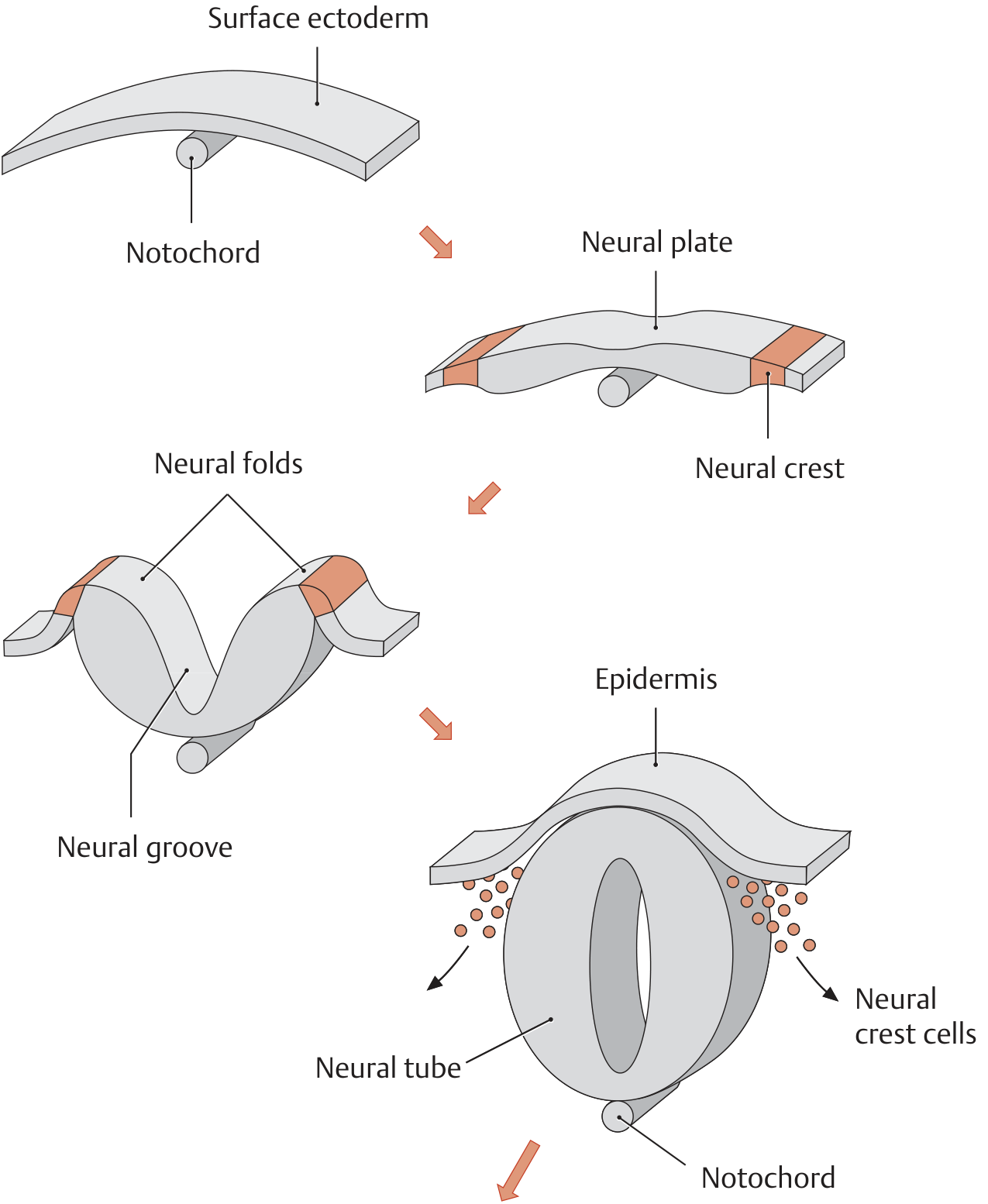
Figure: Sequential formation of the neural crest - from notochord-induced neural plate, through neural folds and groove, to the closed neural tube from which neural crest cells delaminate.
2. Epithelial-to-Mesenchymal Transition (EMT)
Before migrating, NCCs must convert from epithelial to mesenchymal cells - a process triggered primarily by BMPs acting on neural epithelial cells:
- BMPs induce expression of zinc finger transcription factors Snail, Slug, and Twist, which have a conserved role in promoting EMT.
- These transcription factors upregulate cytoskeletal remodeling proteins and matrix metalloproteinases (MMPs) that degrade the basement membrane of the neural tube.
- NCC expression of cadherins changes - cells loosen their adhesive contacts with the neural tube.
- NCCs begin expressing integrins (receptors for laminins and collagens) to adhere to extracellular matrix along peripheral migratory routes.
"Neural crest cell migration does not rely on scaffolding (ie, radial glial cells or preexisting axon tracts) and thus is called free migration. This form of neuronal migration requires significant cytoarchitectural and cell adhesive changes and differs from most of the migratory events in the central nervous system." - Kandel's Principles of Neural Science, 6th Ed., p. 1188
3. Timing of Departure
- Cranial (head) region: NCCs begin to migrate before the neural tube is completely closed.
- Trunk region: Migration is delayed until after neural tube closure is complete.
(THIEME Atlas of Anatomy - General Anatomy)
4. Migratory Pathways
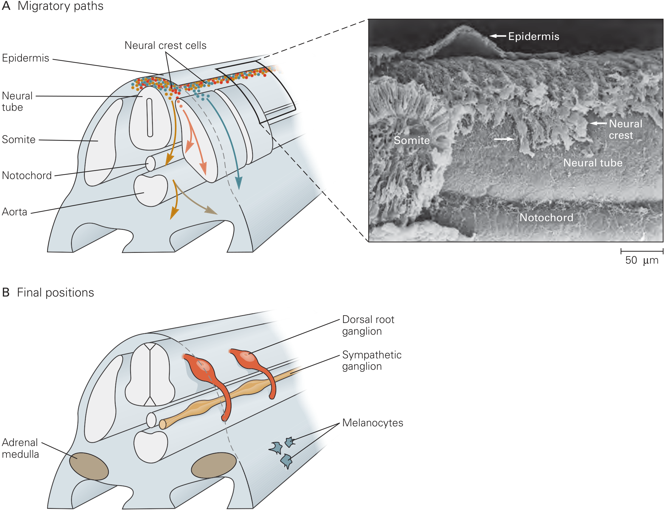
Figure 46-9 from Kandel's Principles of Neural Science: (A) Migratory paths of neural crest cells through the embryo, with SEM showing crest cells adjacent to neural tube and somites. (B) Final positions of major NCC derivatives - dorsal root ganglia, sympathetic ganglia, adrenal medulla, and melanocytes.
Trunk NCC Migration Routes (three main pathways):
| Pathway | Route | Derivatives |
|---|---|---|
| Dorsal (dorsolateral) | Through the dermis, into skin via holes in basal lamina | Melanocytes (melanoblasts) |
| Ventrolateral | Through anterior half of somite | Sensory neurons of dorsal root ganglia |
| Ventral | Medial/ventral path, alongside dorsal aorta | Sympathetic chain ganglia, adrenal medulla chromaffin cells, enteric ganglia, preaortic ganglia |
Somite Channeling - Segmentation
A key feature of trunk NCC migration is that cells pass through only the anterior (rostral) half of each somite, avoiding the posterior half. This segmented channeling is imposed by ephrin B proteins, concentrated in the posterior half of each somite. Ephrins act as repellant signals that interact with EphB class receptor tyrosine kinases on NCCs, blocking invasion of the posterior sclerotome. This creates the metameric (segmental) arrangement of the dorsal root and sympathetic ganglia.
(Langman's Medical Embryology)
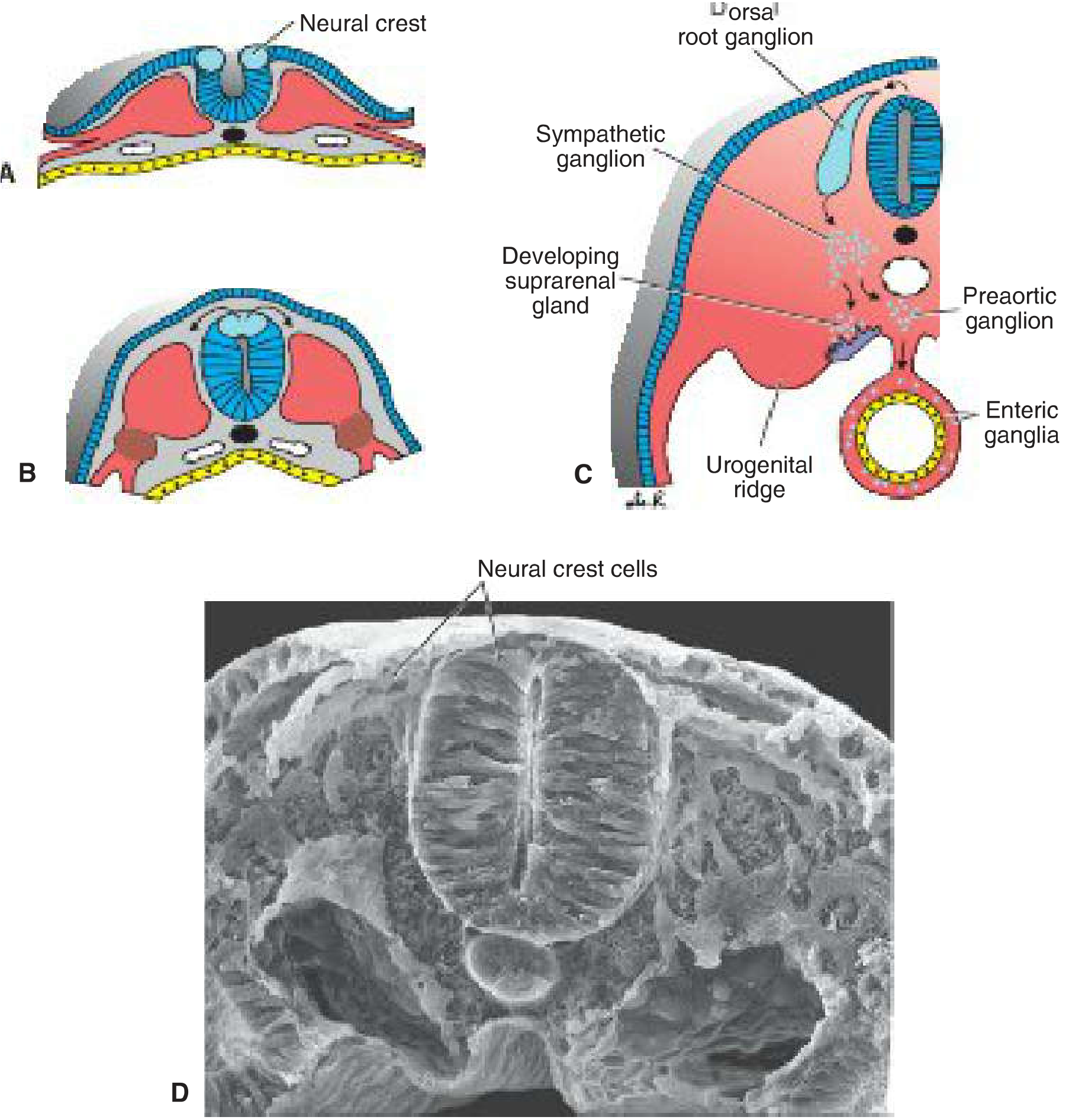
Figure 6.5 from Langman's Medical Embryology: (A-B) Cross-sections showing NCC delamination; (C) Final NCC-derived structures including sympathetic ganglion, developing suprarenal gland, preaortic ganglion, and enteric ganglia; (D) SEM showing neural crest cells on the dorsal surface of the closed neural tube.
5. Guidance Molecules
Several secreted factors promote and guide migration:
| Factor | Role |
|---|---|
| BMPs (from dorsal aorta, later) | Promote EMT and continue to guide ventral migrants; induce Mash1 → sympathetic neuron fate |
| Ephrin B ligands | Repellant; confine migration to anterior somite halves (segmentation) |
| Wnt signals | From dorsal neural tube; induce Neurogenin → sensory neuron fate |
| FGF, TGF-β | Regulate proliferation, migration, and differentiation |
| Laminins/Collagens | ECM components in peripheral pathways; NCCs bind via integrins |
| Semaphorins/Neuropilins | Additional repulsive signals channeling cranial NCCs |
(The Developing Human, p. 210; Kandel's Principles of Neural Science, p. 1188)
6. Fate Determination Along Migration Routes
Differentiation into specific derivatives depends on the combination of signals cells encounter along their journey and intrinsic predispositions that vary along the rostrocaudal axis:
- Sensory neuron fate: Cells within the anterior sclerotome are exposed to Wnt from the dorsal neural tube/somites → express Neurogenin (bHLH TF) → sensory neurons. Further diversification yields nociceptive and proprioceptive subtypes.
- Sympathetic neuron fate: Cells following a medial/ventral path are exposed to BMPs from the dorsal aorta → express Mash1 (bHLH TF) → sympathetic neurons.
- Melanocyte fate: Cells taking the dorsolateral path into the epidermis → melanoblasts → melanocytes in skin and hair follicles.
7. Neural Crest Derivatives (Summary Table)
From Langman's Medical Embryology (Table 6.1):
Nervous system:
- Spinal (dorsal root) ganglia
- Sympathetic chain and preaortic ganglia
- Parasympathetic ganglia of the GI tract (enteric ganglia)
- Cranial nerve ganglia (CN V, VII, IX, X - partially)
- Schwann cells and glial cells
- Meninges (forebrain)
- Adrenal medulla (chromaffin cells)
Craniofacial / skeletal:
- Connective tissue and bones of the face and skull
- Odontoblasts (dentin-forming cells)
- Dermis in face and neck
- Smooth muscle to blood vessels of the face and forebrain
Endocrine:
- C cells (parafollicular cells) of the thyroid gland
- Conotruncal septum in the heart
Pigment:
- Melanocytes
8. Clinical Significance
Defective NCC migration or differentiation underlies a broad range of conditions ("neurocristopathies"):
| Disease | NCC Defect |
|---|---|
| Hirschsprung disease | Failure of enteric NCCs to fully colonize the hindgut → aganglionic colon |
| DiGeorge syndrome (22q11.2 deletion) | Defective NCC migration into pharyngeal arches/pouches → absent thymus, parathyroids, conotruncal heart defects |
| Waardenburg syndrome | Mutations in SOX10, PAX3 → melanocyte/cochlear NCC defects → pigmentation + hearing loss |
| CHARGE syndrome | CHD7 mutations → NCC migration defects |
| Treacher Collins syndrome | Cranial NCC apoptosis → craniofacial defects |
| Melanoma, neuroblastoma, pheochromocytoma | NCC-derived tumors; tend to be highly malignant due to the intrinsic migratory/invasive capacity of NCC |
| Neurofibromatosis type 2 (NF2) | Transient change in NF2 promoter activity during NCC migration linked to Schwann cell tumors |
(Fitzpatrick's Dermatology; Langman's Medical Embryology; THIEME Atlas)
"NCC are involved in at least one-third of all birth defects and many cancers such as melanomas, neuroblastomas, and others." - Langman's Medical Embryology
Sources: Kandel's Principles of Neural Science 6th Ed., p. 1188 | Langman's Medical Embryology | The Developing Human: Clinically Oriented Embryology | THIEME Atlas of Anatomy - General Anatomy | Fitzpatrick's Dermatology
This is a shared conversation. Sign in to Orris to start your own chat.