
Tongue Anatomy with reference to Vishram Singh book
tongue anatomy dorsal surface papillae diagram
Vishram Singh Textbook of Anatomy Head Neck Brain tongue anatomy papillae muscles nerve supply
https://notesmed.com/wp-content/uploads/2021/11/Vishram-Sing…
tongue papillae circumvallate fungiform filiform
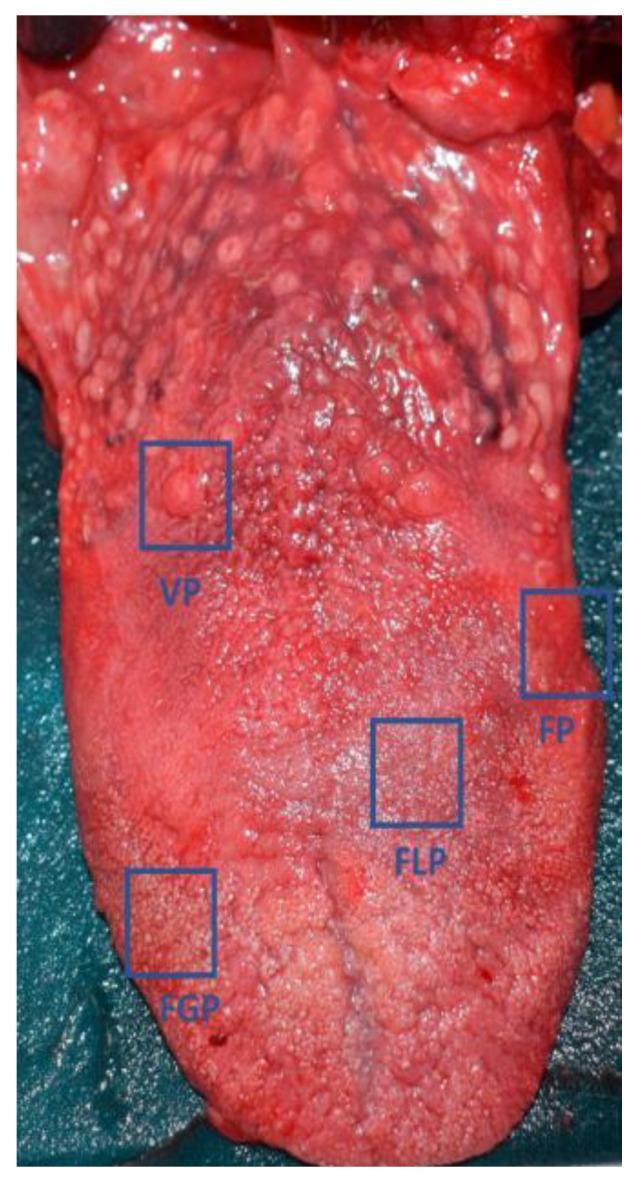
A clinical photograph of a human cadaver tongue, providing a macroscopic view of the dorsal and lateral surfaces to demonstrate the distribution of lingual papillae. The image features blue bounding boxes and labels identifying the four primary types of papillae: Circumvallate papillae (VP), shown as large, circular, dome-shaped structures arranged in a V-shaped row at the posterior aspect of the tongue; Foliate papillae (FP), located as vertical folds or ridges along the posterolateral margins; Fungiform papillae (FGP), visible as small, reddish, mushroom-shaped dots scattered primarily across the anterior two-thirds and tip; and Filiform papillae (FLP), which are the most numerous, giving the tongue its characteristic rough, grayish-pink texture. The specimen displays a natural reddish-pink color with no obvious gross pathological lesions or significant structural alterations. This image is intended for anatomical education and as a baseline for clinical comparison in studies involving gustatory changes or inflammatory conditions affecting the oral mucosa.
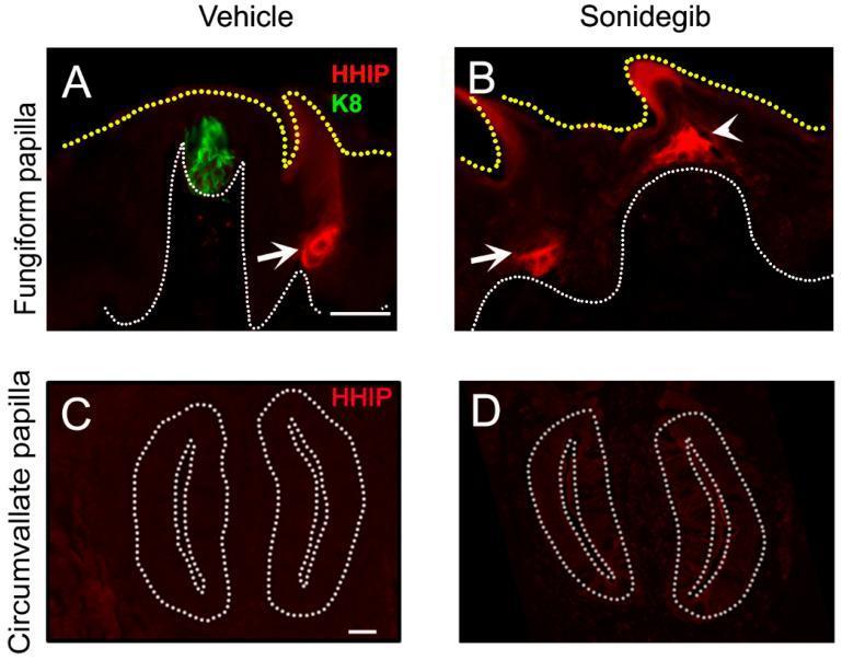
Fluorescence microscopy panels demonstrating Hedgehog (HH) antagonist HHIP expression (red) in lingual papillae under vehicle and Sonidegib (SMO inhibitor) treatment. Panels A and B focus on fungiform papillae (FGP). In the vehicle control (A), HHIP is localized to adjacent filiform papillae (arrow) but absent from the FGP and its K8-positive taste bud (green). After Sonidegib treatment (B), the FGP exhibits a conical morphological shift and ectopic HHIP expression at its apex (arrowhead), while filiform expression (arrow) persists. Panels C and D show circumvallate papillae (CVP). Unlike FGP, the CVP shows negligible HHIP expression in both vehicle (C) and Sonidegib (D) conditions, with no ectopic induction observed following HH pathway inhibition. White dotted lines demarcate the basal lamina/epithelial boundaries, and yellow dotted lines indicate the tongue surface. This comparison illustrates differential molecular and structural responses to HH pathway blockade between anterior (FGP) and posterior (CVP) taste organs, highlighting tissue-specific regulation of HH antagonism.
Tongue Anatomy (Vishram Singh, Textbook of Anatomy: Head, Neck and Brain)
1. External Features (p. 189)
- Root - attached posteriorly to the hyoid bone, mandible, and soft palate
- Body - divided into a dorsal and inferior surface, further split by a V-shaped sulcus terminalis into an anterior two-thirds (oral/palatine part) and posterior one-third (pharyngeal part)
- Apex (tip) - the free anterior end
- Foramen cecum - a small pit at the apex of the sulcus terminalis marking the site of origin of the thyroglossal duct (developmental remnant of the thyroid gland's descent)
- Median sulcus - a shallow midline groove on the dorsum corresponding to the internal fibrous septum that divides the tongue musculature into right and left halves
2. Papillae of the Tongue (p. 190)
| Papilla | Distribution | Feature |
|---|---|---|
| Filiform | Densely cover the entire dorsum | Thread-like, most numerous, purely mechanical (no taste buds) |
| Fungiform | Scattered among filiform, mainly tip and margins | Mushroom-shaped, red dots, contain taste buds |
| Vallate (circumvallate) | 8-12 arranged just anterior to sulcus terminalis, in a V-shaped row | Largest papillae, surrounded by a circular trench; associated von Ebner's serous glands wash the trench |
| Foliate | Posterolateral margins | Vertical mucosal folds, taste buds (rudimentary in humans) |
3. Muscles of the Tongue (p. 191)
- Genioglossus - fan-shaped, from mandible; protrudes the tongue (most important - paralysis causes tongue to fall back and obstruct airway)
- Hyoglossus - from hyoid bone; depresses the tongue
- Styloglossus - from styloid process; retracts and elevates the tongue
- Palatoglossus - from palatine aponeurosis; elevates the tongue root / narrows the oropharyngeal isthmus
4. Nerve Supply (p. 195)
- Motor supply: All muscles (intrinsic and extrinsic) of the tongue are supplied by the hypoglossal nerve (CN XII), except palatoglossus, which is supplied by the cranial root of the accessory nerve via the pharyngeal plexus.
- Sensory supply:
- Anterior two-thirds: general sensation by the lingual nerve (branch of mandibular division of CN V); special taste sensation by the chorda tympani (branch of facial nerve, CN VII)
- Posterior one-third: both general sensation and taste by the glossopharyngeal nerve (CN IX)
- Posteriormost part (near epiglottis): internal laryngeal nerve (branch of the vagus, CN X)
5. Blood Supply and Lymphatic Drainage (p. 194)
- Arterial supply: chiefly the lingual artery (branch of the external carotid artery), supplemented by the tonsillar branch of the facial artery and the ascending pharyngeal artery.
- Venous drainage: via the lingual vein into the internal jugular vein.
- Lymphatic drainage: follows a well-known clinical pattern -
- Tip -> submental nodes (bilateral)
- Anterior two-thirds (excluding tip) -> submandibular nodes
- Posterior one-third -> deep cervical (jugulodigastric) nodes bilaterally directly
6. Development (p. 196)
- Anterior two-thirds: from two lingual swellings and the tuberculum impar of the first arch
- Posterior one-third: from the hypobranchial eminence, contributed to by the third arch (largely overgrowing the second arch contribution) and part of the fourth arch
| Tongue region | Embryonic source | Nerve supply |
|---|---|---|
| Muscles | Occipital myotomes | Hypoglossal (XII) |
| Mucosa, anterior 2/3 | First arch | Lingual (general) + chorda tympani (taste) |
| Mucosa, posterior 1/3 | Third arch | Glossopharyngeal (IX) |
| Mucosa, posteriormost part | Fourth arch | Internal laryngeal (X) |
7. Clinical Correlations (pp. 191, 198)
- Ankyloglossia (tongue-tie): an abnormally short lingual frenulum restricts tongue movement, mentioned by Vishram Singh alongside the description of the frenulum.
- Tongue cancer: most common on the lateral border of the anterior two-thirds; because of the differing lymphatic drainage of anterior vs posterior tongue, the site of a lesion predicts likely nodal spread (submandibular/submental for anterior lesions, bilateral deep cervical for posterior/base lesions) - clinically important for staging and neck dissection planning.
- Hypoglossal nerve palsy: tongue deviates toward the paralyzed side on protrusion (due to unopposed action of the healthy genioglossus) and shows ipsilateral wasting/fasciculation.