
"idiopathic pulmonary fibrosis" AND "usual interstitial pneumonia" AND diagnosis
HRCT chest UIP usual interstitial pneumonia honeycombing subpleural basal fibrosis
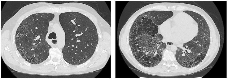
Two axial slices of high-resolution computed tomography (HRCT) of the chest demonstrating a classic Usual Interstitial Pneumonia (UIP) pattern. The images show significant bilateral lung parenchymal abnormalities with a subpleural and basal predominance. Key radiologic features include extensive honeycombing, characterized by clustered, thick-walled subpleural cystic airspaces, and prominent reticular opacities. Additionally, there is evidence of traction bronchiectasis, where the fibrotic destruction of the lung architecture has caused irregular dilation of the bronchi. These findings are consistent with end-stage pulmonary fibrosis, often seen in idiopathic pulmonary fibrosis or connective tissue disease-associated interstitial lung disease (such as systemic sclerosis). The images serve as an educational example of advanced architectural distortion and the characteristic distribution of UIP-pattern fibrosis, distinguishing it from other interstitial lung diseases by the presence of honeycombing and peripheral gradient.
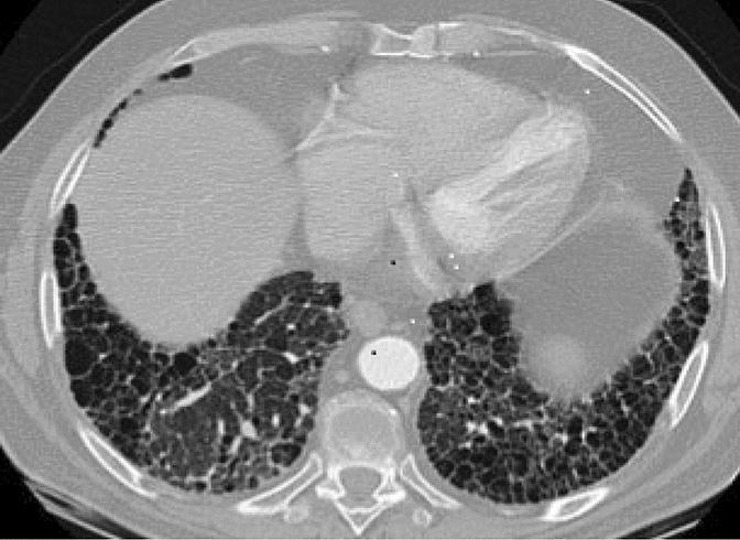
This transverse high-resolution computed tomography (HRCT) axial scan of the chest demonstrates a classic usual interstitial pneumonia (UIP) pattern, frequently associated with fibrotic interstitial lung disease (fILD) in patients with rheumatoid arthritis. The imaging highlights significant architectural distortion characterized by extensive honeycombing, which appears as clustered cystic air spaces with thick, well-defined walls. These abnormalities show a clear basal and subpleural predominance, extending toward the lung periphery while relatively sparing the more central parenchymal regions. Prominent reticular opacities are intermixed with the cystic changes, indicating chronic pulmonary fibrosis. This visual presentation is key for differentiating insidious rheumatoid arthritis-related fibrotic lung disease from acute inflammatory conditions like methotrexate-induced pneumonitis, which typically presents with diffuse ground-glass opacities rather than the honeycombing and structural remodeling shown here.
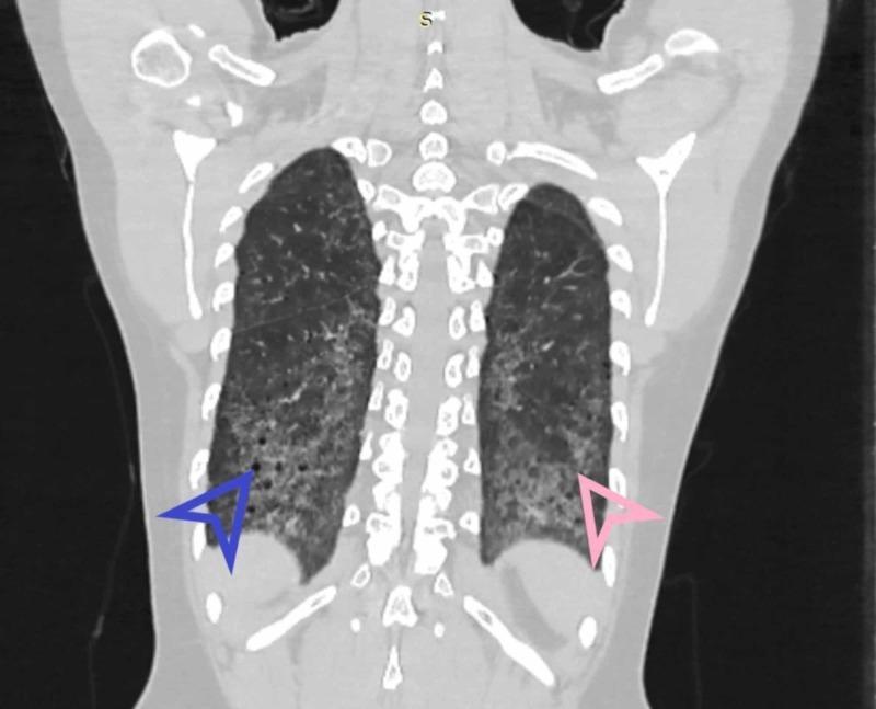
This diagnostic image is a coronal High-Resolution Computed Tomography (HRCT) scan of the chest, focusing on the lung parenchyma. The image demonstrates classic radiological features of Usual Interstitial Pneumonia (UIP), characterized by a marked basal and peripheral predominance of interstitial lung disease. Key findings include honeycombing, indicated by a blue arrowhead in the left lower lobe, appearing as clustered, thick-walled subpleural cystic airspaces. Fibrosis is also highlighted by a pink arrowhead in the right lower lobe, manifesting as reticular opacities and architectural distortion. The distribution is bilateral and symmetric, with the most severe changes occurring at the lung bases. This visual representation is essential for the diagnosis of chronic fibrosing interstitial pneumonias and illustrates the hallmark features used to differentiate UIP from other patterns like Nonspecific Interstitial Pneumonia (NSIP).
CT Chest Analysis - Mr. Munna Lal Pathak (Polaris Neuroscience Hospital, Agra)
Important disclaimer: This is an educational AI-assisted image analysis only. It is not a formal radiology report and cannot substitute for official interpretation by a qualified radiologist. All findings must be correlated with clinical history, pulmonary function tests, and formal DICOM workstation review.
Study Components
- Film 1: Coronal and sagittal multiplanar reformats (MPR) + 3D volume rendering
- Film 2: Axial HRCT slices in lung window (covering upper, mid, and lower chest)
- Film 3: CT thorax plain with soft tissue window; axial slices from neck/thoracic inlet through upper abdomen; includes chest radiograph localizers
Key CT Findings
1. Lung Parenchyma (Dominant Finding)
| Feature | Description |
|---|---|
| Honeycombing | Prominent subpleural clustered cystic airspaces (3-10 mm) with thick, shared walls arranged in multiple layers - most severe in bilateral lower lobes |
| Reticular opacities | Coarse, bilateral intralobular and interlobular septal thickening throughout both lungs |
| Traction bronchiectasis | Irregular, dilated bronchi pulled open by surrounding fibrotic tissue - well seen in lower lobes |
| Distribution | Bilateral, symmetric, subpleural and basal predominant - characteristic gradient from apex (less affected) to base (severely affected) |
| Architecture | Severe architectural distortion; near-total effacement of normal secondary lobular anatomy in lower lobes |
| Lung volume | Significant bilateral lung volume loss (restrictive physiology expected) |
2. Ground Glass Opacity
- Fine intralobular septal thickening below CT resolution (pseudo-GGO)
- Possibly a superimposed acute exacerbation component (though no dominant pure-GGO areas are seen)
3. Nodules / Masses
4. Pleural Disease
- No significant pleural effusion bilaterally
- Mild irregular pleural thickening (reactive, secondary to adjacent severe subpleural honeycombing)
- The characteristic "shaggy" or bumpy pleural surface is well demonstrated on the 3D volume rendering in Film 1
5. Mediastinum and Lymph Nodes (Film 3 - Soft Tissue Window)
- Mediastinum appears midline; trachea and main bronchi are patent
- The third film (soft tissue/thorax plain window) shows mediastinal structures more clearly; no gross bulky mediastinal mass is evident
- Mild reactive mediastinal/hilar lymphadenopathy (commonly seen in active IPF) cannot be excluded without dedicated lymph node window review
- Upper abdomen (Film 3 lower rows): Liver, spleen, and upper abdominal structures appear grossly unremarkable on available images; no obvious hepatic lesions or ascites identified
6. Volume Rendering (3D, Film 1 bottom row)
- Marked reduction in bilateral lung volumes
- Severely irregular, "cauliflower-like" or bumpy lung surface reflecting end-stage subpleural honeycombing
- Normal smooth lung contour is completely lost
Radiological Pattern Classification
"Honeycomb is a distinctive feature for the radiological definition of usual interstitial pneumonitis (UIP)... The extensive fibrosis accompanying honeycomb is associated with distortion of normal lung morphology, resulting in irregular dilatation of segmental and subsegmental airways (traction bronchiectasis/bronchiolectasis)."
- Grainger & Allison's Diagnostic Radiology
- ✅ Subpleural, basal predominant distribution (may be diffuse)
- ✅ Honeycombing with or without peripheral traction bronchiectasis/bronchiolectasis
- ✅ Bilateral involvement
- ✅ Architectural distortion
Differential Diagnosis
| Condition | Notes |
|---|---|
| Idiopathic Pulmonary Fibrosis (IPF) | Most common cause of UIP in older adults (>60 yrs), male predominance, no identifiable cause |
| CTD-associated ILD | Rheumatoid arthritis, systemic sclerosis, polymyositis/dermatomyositis - check ANA, RF, anti-CCP, anti-Scl-70 |
| Chronic Hypersensitivity Pneumonitis (CHP) | Upper lobe involvement, air trapping, exposure history - less likely if purely basal |
| Drug-induced fibrosis | Amiodarone, methotrexate, nitrofurantoin - check medication history |
| Familial/Genetic ILD | Telomere-related mutations - if family history present |
Recommended Workup
- Pulmonary Function Tests (PFTs): Expect reduced FVC, TLC, DLCO (restrictive + reduced gas transfer)
- Autoimmune serology: ANA, RF, anti-CCP, anti-Scl-70, anti-Jo-1, anti-MDA-5 (to exclude CTD-ILD)
- 6-minute walk test (6MWT): For functional assessment and staging
- Bronchoalveolar lavage (BAL): If needed to exclude eosinophilic or infectious causes
- Echocardiography: Pulmonary hypertension is a common complication of advanced IPF
- Multidisciplinary team (MDT) discussion: Pulmonologist + radiologist + pathologist
Management Implications
- Antifibrotic therapy: Nintedanib or Pirfenidone are the two approved agents that slow disease progression per current ATS guidelines
- Supplemental oxygen for exertional and resting hypoxia
- Pulmonary rehabilitation
- Lung transplantation evaluation for eligible patients with progressive disease
- Avoid IPF triggers: Smoking, occupational exposures, GERD treatment (aspiration)
- Vaccinations: Influenza, COVID-19, pneumococcal
Summary Impression
Advanced bilateral pulmonary fibrosis showing a classic "definite" UIP pattern (bilateral subpleural basal-predominant honeycombing, traction bronchiectasis, architectural distortion, lung volume loss). This is highly consistent with Idiopathic Pulmonary Fibrosis (IPF) or a connective tissue disease-associated ILD. No pleural effusion, no dominant pulmonary mass. Upper abdominal organs appear grossly unremarkable. Formal radiological correlation with DICOM images and clinical history is mandatory for definitive diagnosis.
Patient on ventilator on and off post head injury post decompression surgery
Revised CT Interpretation - Critical Clinical Context: Post-TBI, Post-Decompression, On-and-Off Ventilator
Why the UIP/IPF Interpretation Needs Revision
Most Likely Diagnoses (Ranked by Probability)
1. Ventilator-Associated Pneumonia (VAP) - HIGH PROBABILITY
- Caused by gram-negative organisms (Klebsiella, Pseudomonas, Acinetobacter), gram-positives (MRSA), or polymicrobial
- Produces bilateral infiltrates, reticular opacities, consolidation - matching what is seen
- Acinetobacter VAP specifically produces bilateral infiltrates, pleural effusion, cavitations, and hypoxemia (as noted in Goldman-Cecil Medicine)
- The CT pattern can mimic fibrosis in severe/chronic cases
- Diagnosis: Clinical criteria (fever, purulent secretions, new infiltrates, elevated WBC) + BAL culture + CPIS score
2. Ventilator-Induced Lung Injury (VILI) - HIGH PROBABILITY
"VILI mechanisms include barotrauma, volutrauma (diffuse alveolar injury resulting from overdistension), or atelectrauma (injury caused by repeated cycles of recruitment and derecruitment)... In current ventilators, advanced options that limit tidal volume are used to potentially mitigate these harms."
- Atelectrauma from derecruitment between ventilator episodes
- Volutrauma from uncontrolled high tidal volumes during distress episodes
- Diffuse alveolar damage that progresses to fibrosis-like changes on CT
3. Neurogenic Pulmonary Edema (NPE) + Progression to ARDS
"Neurogenic pulmonary edema can develop minutes to days after head trauma, leading to extravascular fluid in the lungs, hypoxia, and decreased lung compliance. The catecholamine surge that often follows TBI can result in increased intravascular pressure, increased capillary permeability, and hydrostatic edema. Additionally, a systematic inflammatory reaction leads to endothelial damage and vasogenic edema."
| Stage | Event |
|---|---|
| Acute TBI | Catecholamine surge → increased pulmonary capillary pressure |
| Days 1-3 | Neurogenic pulmonary edema → bilateral infiltrates |
| Decompression surgery | Surgical stress + anesthesia → aspiration risk |
| Repeated ventilation | VILI, atelectrauma, VAP superimposition |
| Chronic phase | Diffuse alveolar damage organizing → fibrosis-like CT changes |
4. Aspiration Pneumonitis / Pneumonia
- Bilateral dependent infiltrates (gravity-dependent consolidation)
- Organizing pneumonia pattern
- Progressive fibrotic-appearing changes on CT
5. Hospital-Acquired Pneumonia (HAP) from Multidrug-Resistant Organisms
Key Clinical Distinction: Acquired vs. Pre-existing Disease
| Feature | Pre-existing UIP/IPF | Acquired (VAP/VILI/NPE) |
|---|---|---|
| Age at onset | Usually >60 yrs | Any age |
| Prior CT (before injury) | Would show same pattern | Normal or different |
| Timeline | Gradual, years | Weeks-months of ICU stay |
| Lung biopsy pattern | UIP (temporal uniformity lost) | DAD, organizing pneumonia, BOOP |
| Response to antibiotics | None | Partial/complete (if VAP) |
| PFTs before TBI | Would show restriction | Previously normal expected |
| Autoimmune serology | May be positive (CTD-ILD) | Negative |
The Ventilator Management Challenge in TBI + Lung Injury
"Treatment of acute lung injury in TBI is challenging because traditional treatment measures, including hypercapnia, fluid restriction, and prone ventilation (which raises ICP), are contraindicated in TBI."
| Lung-Protective Strategy | Effect on TBI | Management |
|---|---|---|
| Low tidal volume (6 mL/kg PBW) | Can cause hypercapnia → ↑ICP | Permissive hypercapnia generally contraindicated; target PaCO2 35-45 mmHg |
| High PEEP | Reduces venous return → ↑ICP | Use cautiously with ICP monitoring; PEEP 5-8 cmH2O is generally safe |
| Prone positioning | ↑ICP, dangerous | Avoid unless ICP is controlled/monitored |
| Fluid restriction | Can reduce cerebral perfusion pressure | Must balance CPP vs. pulmonary edema |
| Permissive hypoxemia | Worsens secondary brain injury | Target SpO2 >94%, PaO2 >60-80 mmHg |
Immediate Workup Priorities
- BAL / tracheal aspirate cultures - identify VAP organism, guide antibiotic de-escalation
- Serial ABGs - PaO2/FiO2 ratio to diagnose/grade ARDS (Berlin criteria)
- CXR comparison - compare with any previous films to establish trajectory
- Procalcitonin + CRP + CBC - infectious vs. sterile inflammatory etiology
- Echocardiography - rule out neurogenic cardiomyopathy (Takotsubo) contributing to pulmonary edema
- ICP monitoring - if not already in place, critical for ventilator management decisions
- Serum LDH, D-dimer - if PE contributing to hypoxia
- Review ventilator settings - check current tidal volumes, plateau pressures, driving pressures
Management Summary
- Lung-protective ventilation: tidal volume 6 mL/kg predicted body weight, plateau pressure <30 cmH2O, driving pressure <15 cmH2O
- PEEP titration with ICP monitoring (PEEP 5-8 cmH2O usually tolerated)
- Treat VAP empirically with broad-spectrum antibiotics covering gram-negatives and MRSA (piperacillin-tazobactam or carbapenem + vancomycin/linezolid), then de-escalate based on culture
- Elevate head of bed 30-45 degrees (reduces both VAP and ICP)
- Aggressive chest physiotherapy, bronchoscopic clearance if mucus plugging
- Corticosteroids only if organizing pneumonia/BOOP pattern confirmed (controversial in TBI)
- Maintain CPP 60-70 mmHg
- Target PaCO2 35-45 mmHg (avoid both hyperventilation and hypercapnia)
- Avoid hypoxia at all costs (SpO2 >94%)
- Continue ICP management per neurosurgical team
- Spontaneous breathing trials (SBT) should be coordinated with neurosurgical assessment
- The "on-and-off" pattern suggests repeated failed extubations - consider formal swallowing assessment, tracheostomy if prolonged ventilation anticipated (generally recommended if ventilation >14 days expected)