
Make a pptx on the topics oesteoarthritis around 25 pages and each and every topic sholud be cover of oesteoarthritis
osteoarthritis overview diagnosis management NICE guideline 2023
- Definition and burden of osteoarthritis
- Normal joint cartilage and pathophysiology
- Risk factors and causes
- Commonly affected joints: knee, hip, hand, spine, foot
- Symptoms and signs
- Clinical assessment and differential diagnosis
- Investigations and imaging
- Disease severity and complications
- Non-drug management: education, exercise, physiotherapy, weight management
- Medicines and injections
- Surgical management and joint replacement
- Rehabilitation, prevention, prognosis, and patient case examples
Which emphasis should the 25-slide osteoarthritis presentation use?
- Definition and burden of osteoarthritis
- Normal joint cartilage and pathophysiology
- Risk factors and causes
- Commonly affected joints: knee, hip, hand, spine, foot
- Symptoms and signs
- Clinical assessment and differential diagnosis
- Investigations and imaging
- Disease severity and complications
- Non-drug management: education, exercise, physiotherapy, weight management
- Medicines and injections
- Surgical management and joint replacement
- Rehabilitation, prevention, prognosis, and patient case examples
osteoarthritis AND management
https://www.nice.org.uk/guidance/ng226/chapter/Recommendatio…
~/
osteoarthritis knee radiograph joint space narrowing osteophytes
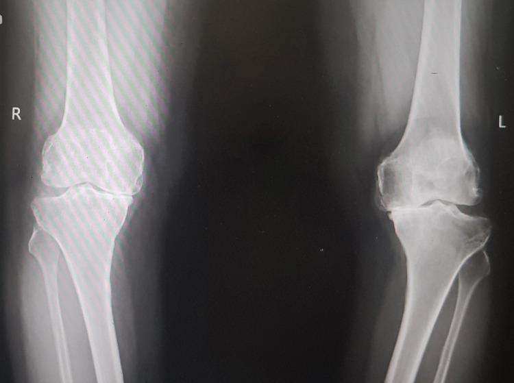
This diagnostic image is an anteroposterior (AP) standing radiograph of bilateral knee joints, demonstrating findings consistent with advanced primary osteoarthritis. The right knee shows significant medial compartment joint space narrowing, subchondral sclerosis, and marginal osteophyte formation at the femoral and tibial condyles. The left knee exhibits even more severe degenerative changes, characterized by near-total loss of medial joint space (bone-on-bone contact), pronounced subchondral sclerosis, large osteophytes, and visible bony irregularity indicating advanced articular cartilage destruction. There is evidence of varus malalignment, particularly on the left. This radiograph serves as a classic educational example of end-stage degenerative joint disease requiring clinical correlation for surgical planning, such as total knee arthroplasty. The image highlights key radiographic hallmarks of osteoarthritis: joint space narrowing, osteophytosis, subchondral sclerosis, and subchondral cyst formation.
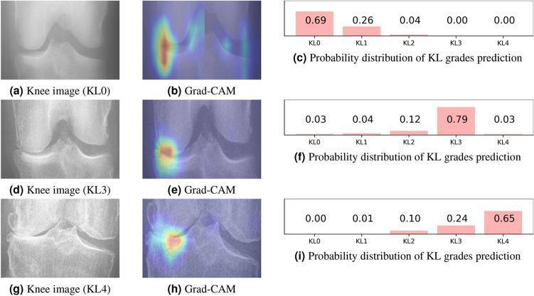
A comparative diagnostic visualization showcasing different stages of knee osteoarthritis using the Kellgren-Lawrence (KL) grading system, integrated with AI explainability heatmaps. The figure is organized into three rows corresponding to KL0 (no OA), KL3 (moderate OA), and KL4 (severe OA). Each row contains a frontal knee radiograph, a Grad-CAM (Gradient-weighted Class Activation Mapping) visualization, and a bar chart of prediction probabilities. Radiographically, KL0 shows a normal joint space and healthy bone contours. KL3 displays definitive joint space narrowing and visible osteophyte formation. KL4 demonstrates severe joint space narrowing with subchondral sclerosis and large, prominent osteophytes. The Grad-CAM overlays indicate the neural network's focus, which shifts from diffuse areas in KL0 to highly localized, intense heatmaps concentrated on the medial joint compartment and marginal osteophytes in the pathological KL3 and KL4 cases. This clinical imaging tool illustrates how computer-aided diagnosis identifies disease markers like joint space loss and bone remodeling to classify osteoarthritis severity.
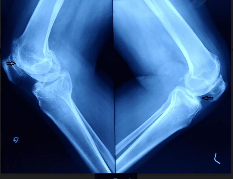
This diagnostic image is a lateral-view plain radiograph of both knee joints showing advanced degenerative changes consistent with Kellgren-Lawrence grade IV osteoarthritis. The imaging demonstrates significant joint space narrowing in the tibiofemoral compartments and evidence of patellofemoral osteoarthritis. Key radiological features include subchondral sclerosis, which appears as increased bone density at the joint margins, and prominent osteophyte formation. Black arrows specifically highlight osteophytes at the superior and inferior poles of the patella and the anterior tibial margins. There is visible narrowing of the patellofemoral joint space. While lateral views typically focus on sagittal alignment, the severe degeneration shown is associated with clinical varus deformity as noted in the patient's history. These findings are representative of end-stage joint disease, which in this clinical context was secondary to ochronotic arthropathy, and indicate the need for total knee arthroplasty (TKA).
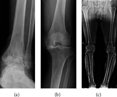
A composite image consisting of three anteroposterior (AP) radiographs demonstrating advanced multi-joint osteoarthritis and lower limb malalignment. Panel (a) shows an AP radiograph of the left ankle with Takakura stage 4 osteoarthritis, characterized by complete joint space loss between the tibia and talus, marginal osteophyte formation, and subchondral sclerosis. Panel (b) illustrates a left knee radiograph with Kellgren-Lawrence Grade IV changes, including severe medial compartment narrowing, osteophytes, and subchondral bone remodeling. Panel (c) is a preoperative standing weight-bearing AP radiograph of the bilateral lower extremities, showing a significant varus deformity of the left leg with a measured hip-knee-ankle (HKA) angle of 14.3 degrees. This series illustrates the clinical correlation between severe distal joint degeneration and global mechanical limb alignment issues in an orthopedic context.
osteoarthritis articular cartilage pathology diagram
mkdir -p "/tmp/workspace/osteoarthritis_ppt/src" "/tmp/workspace/osteoarthritis_ppt/assets" "/tmp/workspace/osteoarthritis_ppt/output" && python /tmp/skills/shared/scripts/fetch_images.py "https://cdn.orris.care/cdss_images/9eca3095a6a7a10ade646ec1abc2b62ab291a9fab6b5512a70acbefb669354ec.png" "https://cdn.orris.care/cdss_images/pmc_clinical_VQA_2bdaa2ef6d66091e1cad57389cd6935cf98f3d92604caf39957c64466b2299d1.jpg"
~/
"osteoarthritis"[MeSH Terms] AND management
NICE NG226 osteoarthritis diagnosis management recommendations topical NSAID exercise weight loss 2026
Make pptx for me on topic oesteoarthritis as you know my name is sahil khan and I am submitting it to dr Sanjay chalani now a best pptx the background sheet of pptx is showing oesteoarthritis images in background and also use x ray and mri images to make pptx more impact full and make last page of thanks you for listening me pptx around 30 with good content according to medical students make border lineing in orange colour and font in black colour write definitions properly from refrance of orthopaedic books but don't mention name of books