
Vertigo
vertigo diagnosis management
vertigo types peripheral central vestibular diagram
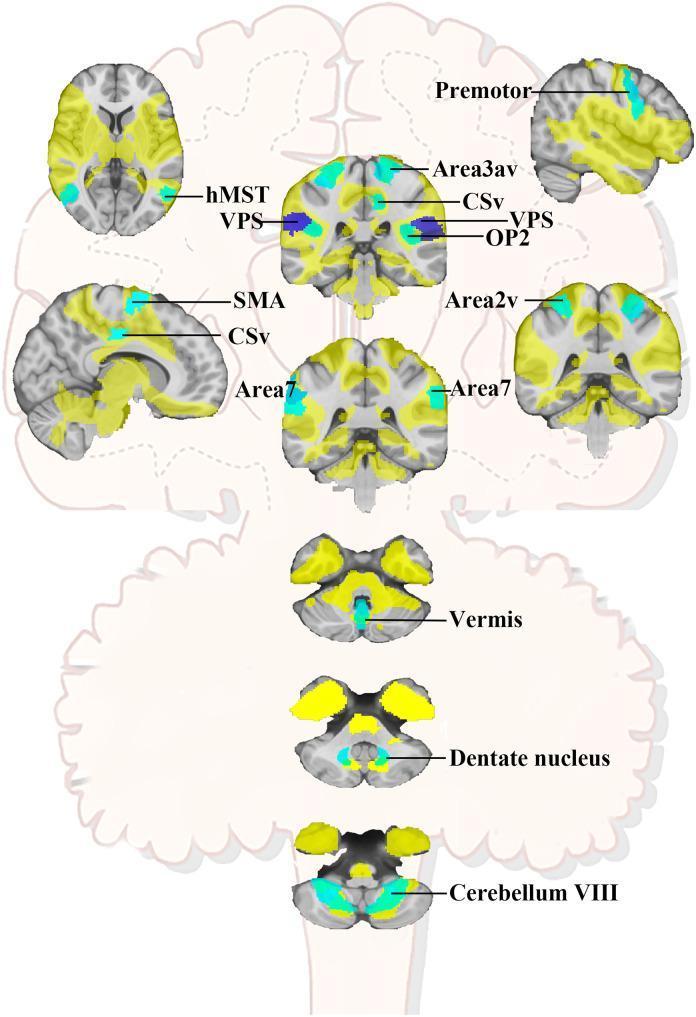
This medical schematic illustration depicts the lesion-based vertigo network across multiple neuroanatomical planes, including axial, sagittal, and coronal views of the brain and cerebellum. The diagram identifies key cortical and subcortical regions involved in vestibular processing and vertigo pathophysiology. Specific regions highlighted in teal and dark blue include the bilateral Area 2v (intraparietal sulcus), Area 3av (central sulcus fundus), Premotor cortex, Cingulate Sulcus Visual area (CSv), Supplementary Motor Area (SMA), and Area 7 (supramarginalis). Critical vestibular hubs such as the Visual Posterior Sylvian (VPS) or Posterior Insular Cortex (PIC) and Parietal Opercula 2 (OP2) are also marked. The lower sections focus on the vestibulocerebellum, highlighting the vermis, bilateral dentate nucleus, and cerebellum lobule VIII. This anatomical map serves as an educational tool for understanding the spatial distribution and connectivity of the functional network associated with acute vertigo following supratentorial or cerebellar lesions.
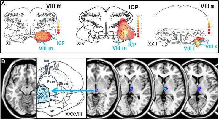
This composite educational image illustrates brainstem lesion locations associated with acute vestibular syndromes. Panel A displays three axial anatomical diagrams of the medulla and pons (levels XII, XIV, XXII) with superimposed heat-map overlays representing infarct overlap in 23 patients with rotational vertigo. The heat maps highlight concentrated involvement in the medial vestibular nucleus (VIII m), superior and lateral vestibular nuclei (VIII s, VIII l), inferior cerebellar peduncle (ICP), and the y-group. A color scale indicates the frequency of lesion overlap. Panel B correlates clinical MRI data with midbrain anatomy (level XXXVIII). It features axial T1-weighted MRI slices showing localized rostral mesencephalic-diencephalic infarcts (highlighted in blue/purple) alongside a detailed anatomical diagram identifying the interstitial nucleus of Cajal (iC), Edinger-Westphal nucleus (EW), and nucleus oculomotorius principalis (IIIpr). The figure demonstrates the anatomical substrate for central vestibular pseudoneuritis, contrasting lower brainstem lesions causing rotational vertigo with rostral midbrain lesions causing stance and gait instability.
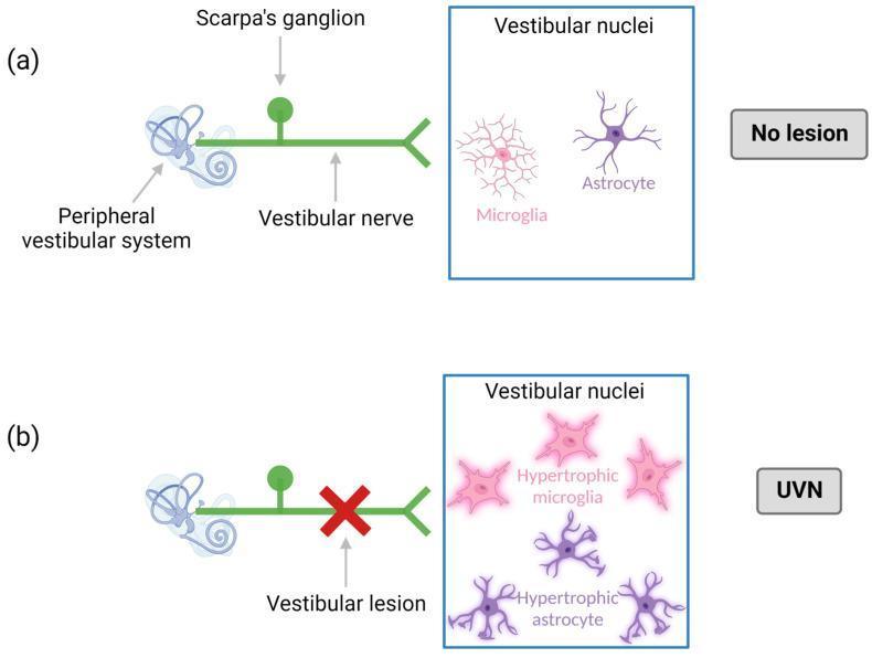
This medical pathophysiology diagram illustrates the neuroinflammatory response in the vestibular system following a unilateral vestibular neurectomy (UVN). The diagram is divided into two panels: (a) 'No lesion' and (b) 'UVN'. Panel (a) depicts a healthy peripheral vestibular system connected to the brainstem via the vestibular nerve and Scarpa's ganglion. Within the vestibular nuclei, microglia and astrocytes are shown in their quiescent state, characterized by small cell bodies and thin, ramified processes. Panel (b) demonstrates the pathophysiological state following a vestibular lesion (marked by a red 'X'). This deafferentation triggers a central inflammatory response within the vestibular nuclei. The diagram shows a proliferation and morphological change in glial cells, specifically highlighting hypertrophic microglia and hypertrophic astrocytes. These cells are depicted with enlarged cell bodies and thickened, more numerous processes, signifying reactive gliosis. This model serves to explain the cellular mechanisms of central compensation and neuroinflammation in acute unilateral peripheral vestibulopathy (AUPV).
Dix-Hallpike maneuver BPPV diagnosis positional test
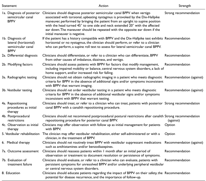
<table> <tr> <th>Statement</th> <th>Action</th> <th>Strength</th> </tr> <tr> <td>1a. Diagnosis of posterior semicircular canal BPPV</td> <td>Clinicians should diagnose posterior semicircular canal BPPV when vertigo associated with torsional, upbeating nystagmus is provoked by the Dix-Hallpike maneuver, performed by bringing the patient from an upright to supine position with the head turned 45° to one side and neck extended 20° with the affected ear down. The maneuver should be repeated with the opposite ear down if the initial maneuver is negative.</td> <td>Strong recommendation</td> </tr> <tr> <td>1b. Diagnosis of lateral (horizontal) semicircular canal BPPV</td> <td>If the patient has a history compatible with BPPV and the Dix-Hallpike test exhibits horizontal or no nystagmus, the clinician should perform, or refer to a clinician who can perform, a supine roll test to assess for lateral semicircular canal BPPV.</td> <td>Recommendation</td> </tr> <tr> <td>2a. Differential diagnosis</td> <td>Clinicians should differentiate, or refer to a clinician who can differentiate, BPPV from other causes of imbalance, dizziness, and vertigo.</td> <td>Recommendation</td> </tr> <tr> <td>2b. Modifying factors</td> <td>Clinicians should assess patients with BPPV for factors that modify management, including impaired mobility or balance, central nervous system disorders, a lack of home support, and/or increased risk for falling.</td> <td>Recommendation</td> </tr> <tr> <td>3a. Radiographic testing</td> <td>Clinicians should not obtain radiographic imaging in a patient who meets diagnostic criteria for BPPV in the absence of additional signs and/or symptoms inconsistent with BPPV that warrant imaging.</td> <td>Recommendation (against)</td> </tr> <tr> <td>3b. Vestibular testing</td> <td>Clinicians should not order vestibular testing in a patient who meets diagnostic criteria for BPPV in the absence of additional vestibular signs and/or symptoms inconsistent with BPPV that warrant testing.</td> <td>Recommendation (against)</td> </tr> <tr> <td>4a. Repositioning procedures as initial therapy</td> <td>Clinicians should treat, or refer to a clinician who can treat, patients with posterior canal BPPV with a canalith repositioning procedure.</td> <td>Strong recommendation</td> </tr> <tr> <td>4b. Postprocedural restrictions</td> <td>Clinicians should not recommend postprocedural postural restrictions after canalith repositioning procedure for posterior canal BPPV.</td> <td>Strong recommendation (against)</td> </tr> <tr> <td>4c. Observation as initial therapy</td> <td>Clinicians may offer observation with follow up as initial management for patients with BPPV.</td> <td>Option</td> </tr> <tr> <td>5. Vestibular rehabilitation</td> <td>The clinician may offer vestibular rehabilitation, either self-administered or with a clinician, in the treatment of BPPV.</td> <td>Option</td> </tr> <tr> <td>6. Medical therapy</td> <td>Clinicians should not routinely treat BPPV with vestibular suppressant medications such as antihistamines and/or benzodiazepines.</td> <td>Recommendation (against)</td> </tr> <tr> <td>7a. Outcome assessment</td> <td>Clinicians should reassess patients within 1 month after an initial period of observation or treatment to document resolution or persistence of symptoms.</td> <td>Recommendation</td> </tr> <tr> <td>7b. Evaluation of treatment failure</td> <td>Clinicians should evaluate, or refer to a clinician who can evaluate, patients with persistent symptoms for unresolved BPPV and/or underlying peripheral vestibular or central nervous system disorders.</td> <td>Recommendation</td> </tr> <tr> <td>8. Education</td> <td>Clinicians should educate patients regarding the impact of BPPV on their safety, the potential for disease recurrence, and the importance of follow-up.</td> <td>Recommendation</td> </tr> </table>
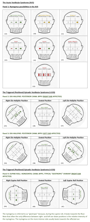
Summary : This figure provides a comprehensive visual guide to nystagmus patterns in acute and triggered (positional) vestibular syndromes, including the acute vestibular syndrome (AVS) and episodic vestibular syndrome (E-VS), with detailed head diagrams illustrating nystagmus direction and affected sides during various diagnostic maneuvers. diagram: # The Acute Vestibular Syndrome (AVS) : • Panel 1: Nystagmus possibilities in the AVS. • Six head diagrams (1A–1F) show different nystagmus directions (horizontal, vertical, torsional, mixed) and their clinical implications. • Diagram 1G summarizes "Central" nystagmus with multidirectional arrows. # The Triggered (Positional) Episodic Vestibular Syndrome (E-VS) : • Panel 2: Dix-Hallpike maneuver for posterior canal BPPV (right ear affected). – Three head diagrams (2A–2C) show nystagmus direction in right Dix-Hallpike, seated, and left Dix-Hallpike positions. • Panel 3: Dix-Hallpike maneuver for posterior canal BPPV (left ear affected). – Three head diagrams (3A–3C) show nystagmus direction in right Dix-Hallpike, seated, and left Dix-Hallpike positions. • Panel 4: Supine roll test for horizontal canal BPPV, "geotropic" variant (right ear affected). – Three head diagrams (4A–4C) show nystagmus direction in right supine roll, seated, and left supine roll positions. # Diagram Features : • Each head diagram is labeled with the maneuver/position and affected side. • Arrows indicate the direction of nystagmus (horizontal, vertical, torsional, or mixed). • Color coding (green, red) highlights affected sides and nystagmus intensity. • Explanatory text beneath each diagram clarifies clinical interpretation. # Analysis : • The diagrams systematically differentiate between central and peripheral causes of nystagmus based on direction, position, and affected side. • The AVS section emphasizes the importance of nystagmus direction (e.g., unidirectional vs. direction-changing) in diagnosis. • The E-VS panels illustrate how nystagmus changes with head position, aiding in the localization of BPPV to specific canals and sides. • The "geotropic" variant is visually distinguished by nystagmus beating towards the floor, with intensity differences between sides.
Vertigo
Definition
Classification: Peripheral vs. Central
| Feature | Peripheral | Central |
|---|---|---|
| Course | Episodic, fatigable | Constant or progressive |
| Onset | Abrupt | Gradual or abrupt |
| Severity | Intense | Milder but persistent |
| Nystagmus | Horizontal or rotary, suppressed by fixation | May be vertical; not suppressed by fixation |
| Neurologic symptoms | Absent | Present (dysarthria, diplopia, ataxia, weakness, numbness) |
| Hearing loss/tinnitus | May be present | Uncommon |
| Head impulse test | Abnormal (catch-up saccade) | Normal |
Differential Diagnosis - Classified by Duration
Peripheral Causes
- Benign Paroxysmal Positional Vertigo (BPPV) - most common cause; brief episodes (<30 s) triggered by head position change; due to canalith (otolith debris) in the posterior semicircular canal; positive Dix-Hallpike test
- Vestibular Neuritis / Labyrinthitis - sudden onset, severe, lasting days to weeks; often follows viral illness; hearing loss present only in labyrinthitis; positive head impulse test
- Meniere Disease - recurrent attacks lasting hours; classic triad of severe rotational vertigo + tinnitus + sensorineural hearing loss; due to endolymphatic hydrops
- Acoustic Neuroma (Vestibular Schwannoma) - gradual unilateral hearing loss and tinnitus; MRI required
- Others: perilymphatic fistula, acute otitis media, labyrinth concussion, motion sickness
Central Causes
- Vertebrobasilar Insufficiency (VBI) / Stroke - in older adults with vascular risk factors; associated neurological symptoms (dysarthria, ataxia, diplopia, weakness); consider posterior circulation TIA
- Cerebellar Hemorrhage/Infarction - sudden severe symptoms; headache, vomiting, ataxia, dysmetria; emergency
- Wallenberg Syndrome (PICA occlusion) - vertigo + loss of pain/temperature (crossed pattern) + ipsilateral Horner syndrome + dysphasia
- Vestibular Migraine - recurrent vertigo episodes associated with migraine headaches; most common central cause in younger patients
- Multiple Sclerosis - vertigo can be the presenting symptom; MRI white matter plaques
- Head trauma, tumors, encephalitis, temporal lobe epilepsy
Key Diagnostic Steps
History (accounts for 85% of diagnosis)
- Character, duration, onset, triggers, positional dependence
- Associated ear symptoms (hearing loss, tinnitus, fullness)
- Neurological symptoms (headache, diplopia, dysarthria, limb weakness)
- Medications with vestibulotoxicity (aminoglycosides, anticonvulsants, quinine, minocycline)
- Vascular risk factors for stroke
Physical Examination
- Dix-Hallpike maneuver - from sitting to supine with head turned 45° to one side; look for torsional upbeating nystagmus (BPPV posterior canal)
- Supine roll test - for horizontal canal BPPV (geotropic nystagmus)
- HINTS exam (for constant/acute vestibular syndrome):
- Head Impulse Test - abnormal = peripheral (catch-up saccade); normal = central (concerning)
- Nystagmus - unidirectional = peripheral; direction-changing or vertical = central
- Test of Skew - present = central
- Orthostatic BP, full cranial nerve exam, Romberg, tandem gait
- Tuning fork tests (Weber, Rinne)
Important: HINTS and Dix-Hallpike should NOT be routinely combined - HINTS is for acute vestibular syndrome (constant vertigo), while Dix-Hallpike is for episodic/positional vertigo. - Rosen's Emergency Medicine
BPPV Clinical Guidelines (strong recommendations):
- Diagnose posterior canal BPPV when Dix-Hallpike provokes torsional upbeating nystagmus
- For horizontal nystagmus or negative Dix-Hallpike, perform supine roll test (lateral canal BPPV)
- Do NOT obtain routine imaging if BPPV criteria are met
- Do NOT order routine vestibular testing for straightforward BPPV
Investigations
- Routine labs: largely unhelpful except finger-stick glucose; CBC/chemistry if anemia or dehydration suspected
- Audiogram: if unilateral otologic symptoms or unexplained vertigo
- Electronystagmography (ENG): objective vestibular localization
- MRI brain: indicated for unilateral otologic symptoms, suspected posterior fossa lesion (acoustic neuroma, MS, stroke), or treatment-resistant vertigo - MRI superior to CT for posterior fossa
- CT head: emergent if cerebellar hemorrhage or infarction is suspected
Treatment
BPPV
- Epley (canalith repositioning) maneuver - first-line for posterior canal BPPV; highly effective
- "Barbecue roll" (Lempert maneuver) - for horizontal canal BPPV; sequential 90° rotations
- Gufoni maneuver - alternative for horizontal canal variant
- No post-procedural postural restrictions needed (strong recommendation against them)
- Vestibular suppressants should NOT be used routinely for BPPV
Vestibular Neuritis / Labyrinthitis
- Supportive care (rest, hydration)
- Vestibular suppressants for acute symptoms
- Corticosteroids (methylprednisolone) - may speed recovery of vestibular function
- Vestibular rehabilitation exercises once acute phase resolves
Vestibular Suppressants (for acute symptomatic relief)
- IV ondansetron 4 mg - recommended first-line IV agent in ED
- Promethazine (Phenergan) - effective but FDA boxed warning for IV use; prefer IM/oral
- Meclizine, dimenhydrinate, lorazepam - used for outpatient management
- These treat symptoms; they do not address the underlying cause
Meniere Disease
- Low-sodium diet, diuretics (hydrochlorothiazide/triamterene)
- Intratympanic gentamicin or steroids
- Endolymphatic shunt surgery for refractory cases
Central Causes
- Stroke/VBI - urgent neurology/stroke team involvement; thrombolytics if eligible, antiplatelets, risk factor control
- Vestibular migraine - migraine prophylaxis (topiramate, valproate, beta-blockers, amitriptyline) and acute migraine treatment
- MS - disease-modifying therapy
Red Flags (Central Vertigo Alarm Features)
- New severe headache
- Diplopia, dysarthria, dysphagia
- Limb ataxia or weakness
- Vertical nystagmus or direction-changing nystagmus
- Normal head impulse test with nystagmus
- Skew deviation
- Inability to walk or stand
- Older age with cardiovascular risk factors and new isolated vertigo
Explain with images Explain with images
inner ear anatomy semicircular canals vestibular labyrinth
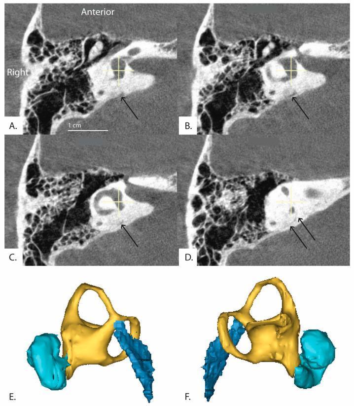
This composite educational resource features high-resolution imaging of the human inner ear anatomy. Panels A-D display sequential axial CT images reformatted in the plane of the horizontal semicircular canal, progressing from an inferior to superior perspective. Black arrows highlight the distal vestibular aqueduct as it traverses the petrous temporal bone. The images demonstrate the spatial relationship between the bony labyrinth, including the vestibule and semicircular canals, and the vestibular aqueduct. Panels E and F provide 3D anatomical reconstructions (right and left views, respectively) illustrating the complex spatial morphology of these structures. The color-coding in the 3D models differentiates key components: the cochlea is shown in cyan, the vestibule and semicircular canals in yellow, and the vestibular aqueduct in dark blue. This visualization is intended to assist in the clinical assessment of inner ear pathologies, such as Meniere's disease or superior canal dehiscence syndrome, by mapping the trajectory and angle of the vestibular aqueduct relative to the labyrinthine structures.
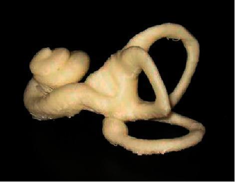
This image presents a 3D computational reconstruction of the human inner ear, derived from a statistical shape model. The anatomical diagram demonstrates the complex three-dimensional relationship between the cochlea, vestibule, and semicircular canals. On the left, the spiral-shaped cochlea is clearly visible with its characteristic bulbous, snail-like winding. This transitions into the central vestibule, from which the three semicircular canals (superior, posterior, and lateral) originate. These canals are depicted as arched, looped structures oriented in nearly orthogonal planes, illustrating their physiological role in sensing rotational head movements. The model features a pale, textured surface, representing the bony labyrinth, set against a high-contrast black background. This visual serves as an educational tool for understanding otolaryngology and vestibular anatomy, particularly for medical students and clinicians studying diagnostic imaging segmentation or the pathophysiology of inner ear disorders like Meniere's disease.
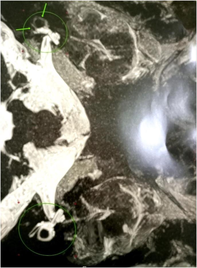
This diagnostic image is a high-resolution grayscale scan of the inner ear, likely a specialized MRI sequence focused on the temporal bone. It illustrates the anatomical structures of the vestibular system, specifically the semicircular canals. The image highlights pathological changes characterized by focal obliteration of the semicircular canal lumens. Instead of the typical smooth, continuous, and rounded contours of a healthy labyrinth, the affected canals exhibit a distinct 'spiky' appearance and irregular margins, as indicated by the green circles and arrows. These visual features are consistent with labyrinthitis ossificans, a process where the fluid-filled spaces of the inner ear are replaced by fibrous tissue or new bone formation following an inflammatory event. This finding is clinically significant for diagnosing causes of sensorineural hearing loss and vestibular dysfunction. The contrast between the bright signal of the remaining fluid-filled spaces and the dark, irregular areas of obliteration allows for the assessment of disease progression within the bony labyrinth.
Epley maneuver canalith repositioning BPPV treatment steps
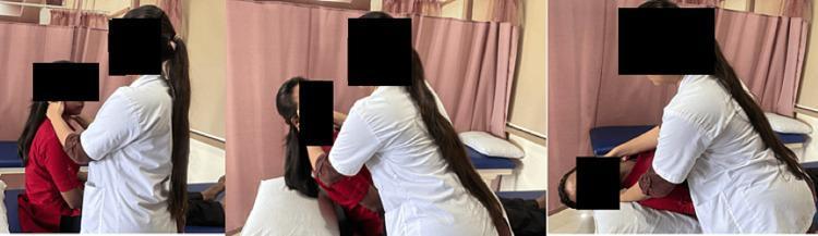
A sequence of three clinical photographs demonstrating the initial phases of the Epley maneuver, a canalith repositioning procedure used to treat benign paroxysmal positional vertigo (BPPV). The images show a healthcare provider in a white clinical coat and a patient on an examination bed in a physiotherapy or neurology setting. In the first panel, the patient is seated upright while the provider stabilizes the head. In the second panel, the provider rotates the patient's head approximately 45 degrees toward the affected side (right). In the third panel, the patient is moved rapidly into a supine position with the head extending off the edge of the supporting pillow to maintain neck extension and rotation. This clinical demonstration illustrates the manual guidance required to facilitate the movement of otoconia within the semicircular canals. The background shows standard clinical equipment including an examination table with blue padding and privacy curtains.
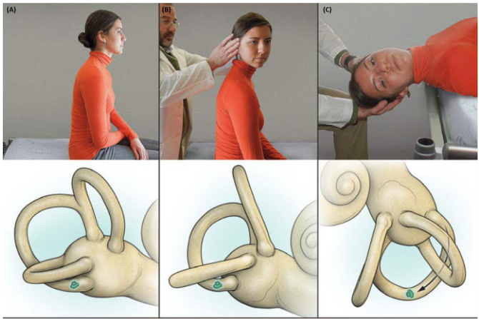
Summary : This figure demonstrates a clinical maneuver for repositioning otoliths in the inner ear, likely related to the treatment of benign paroxysmal positional vertigo (BPPV). The top row shows a sequence of photographs of a patient being guided through the maneuver, while the bottom row provides corresponding anatomical illustrations of the inner ear, highlighting the movement of otoliths. photo and illustration: # Scene Overview : • Top row: Three sequential photographs of a patient in an orange shirt being guided by a clinician through a head movement maneuver. • Bottom row: Three anatomical illustrations of the inner ear, each corresponding to the photograph above, showing the position and movement of otoliths (depicted as green shapes). # Technical Details : • No scale bars or magnification indicated. • The anatomical illustrations focus on the semicircular canals and otoliths, with the cochlea visible in the third panel. # Spatial Relationships : • Left panel (A): Patient is seated upright, head in neutral position; otolith is located in the posterior semicircular canal. • Middle panel (B): Patient’s head is turned 45 degrees to the right by the clinician; otolith remains in the canal. • Right panel (C): Patient is laid back with head extended and rotated; otolith is shown moving through the canal toward the vestibule. # Analysis : • The sequence visually explains the steps of a canalith repositioning maneuver, likely the Epley maneuver, used to treat BPPV by moving displaced otoliths out of the semicircular canal and back into the vestibule. • The anatomical illustrations clarify the intended movement of the otoliths during each step, supporting the clinical photographs. • The figure effectively links clinical technique with underlying anatomical changes.
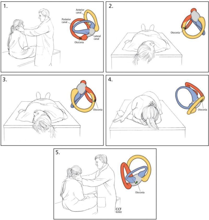
A five-panel medical illustration sequence detailing the Epley maneuver for treating Benign Paroxysmal Positional Vertigo (BPPV). Each panel pairs a clinical illustration of a patient and clinician with a corresponding anatomical diagram of the right inner ear's semicircular canals (anterior in yellow, posterior in red, and lateral in blue). Step 1 shows the patient seated with the head rotated 45° toward the affected side, with otoconia located in the posterior canal. Step 2 depicts the patient moved into a supine position with the neck slightly extended. Step 3 illustrates the head being rotated 90° toward the opposite (healthy) side. Step 4 shows the patient rolling onto their side while maintaining head rotation, facilitating the movement of otoconia through the canal toward the utricle. Step 5 shows the patient returning to a seated position. The anatomical insets track the gravitational migration of otoconia out of the posterior semicircular canal, demonstrating the mechanical rationale for this canalith repositioning procedure.
Meniere disease endolymphatic hydrops inner ear pathology
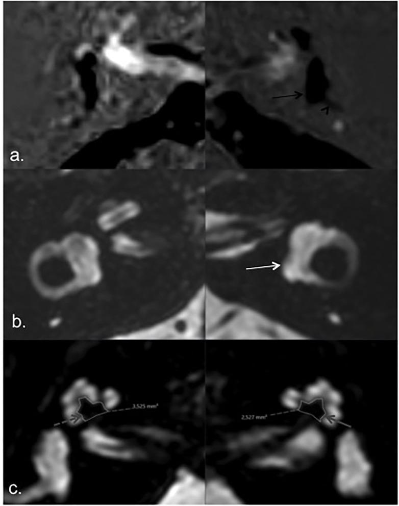
Multi-panel axial Magnetic Resonance Imaging (MRI) study of the inner ear focusing on Ménière’s disease pathology. Panel (a) displays HYDROPS sequences showing bilateral vestibular endolymphatic hydrops, with more severe distension on the left side (black arrow) and herniation of the utricle into the non-ampullary limb of the lateral semicircular canal (black arrowhead). Panel (b) presents axial T2-weighted images highlighting the total membranous labyrinth (white arrow). Panel (c) shows comparative axial T2 images focused on the cochlear modiolus area. The images demonstrate significant asymmetry between the 'index' (symptomatic) left ear and the asymptomatic right ear; the modiolar area on the left (solid gray arrow) is markedly smaller (2.327 mm²) compared to the right (3.525 mm², dashed gray arrow). This diagnostic imaging illustrates the relationship between endolymphatic hydrops and structural changes in the cochlear modiolus, relevant for otolaryngology and neurotology educational purposes.
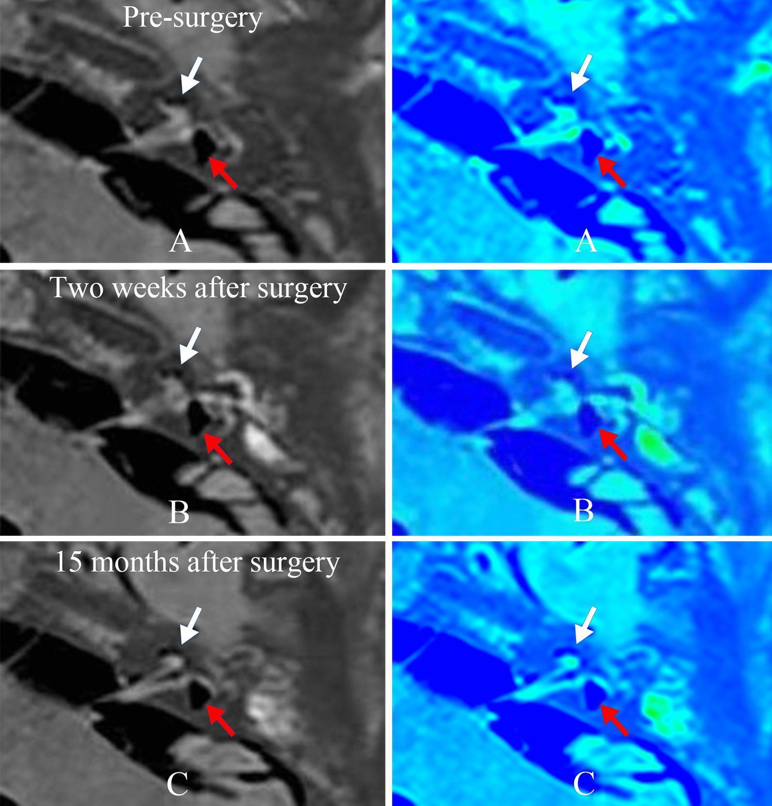
Diagnostic axial MRI scans of the inner ear illustrating endolymphatic hydrops (EH) in a patient with Ménière’s disease across three time intervals: pre-surgery (A), two weeks post-surgery (B), and 15 months post-surgery (C). The left column displays grayscale gadolinium-enhanced MRI (Gd-MRI) using a three-dimensional real inversion recovery sequence. White arrows indicate the cochlea, and red arrows indicate the vestibule. The right column provides corresponding pseudocolor images where blue represents the endolymphatic space and green represents the perilymphatic space. In the pre-surgery baseline (A), significant cochlear and vestibular hydrops are visible, characterized by the expansion of the endolymphatic (blue) compartments at the expense of the perilymphatic (green) areas. Sequential follow-up images (B and C) demonstrate a consistent appearance with no significant reduction or change in the degree of hydrops following endolymphatic sac decompression (ESD) surgery. This comparison illustrates a case where surgical intervention did not result in visual anatomical reversal of fluid compartment distention, serving as a radiological reference for post-operative monitoring of inner ear pathology.
HINTS exam head impulse test nystagmus skew deviation vertigo
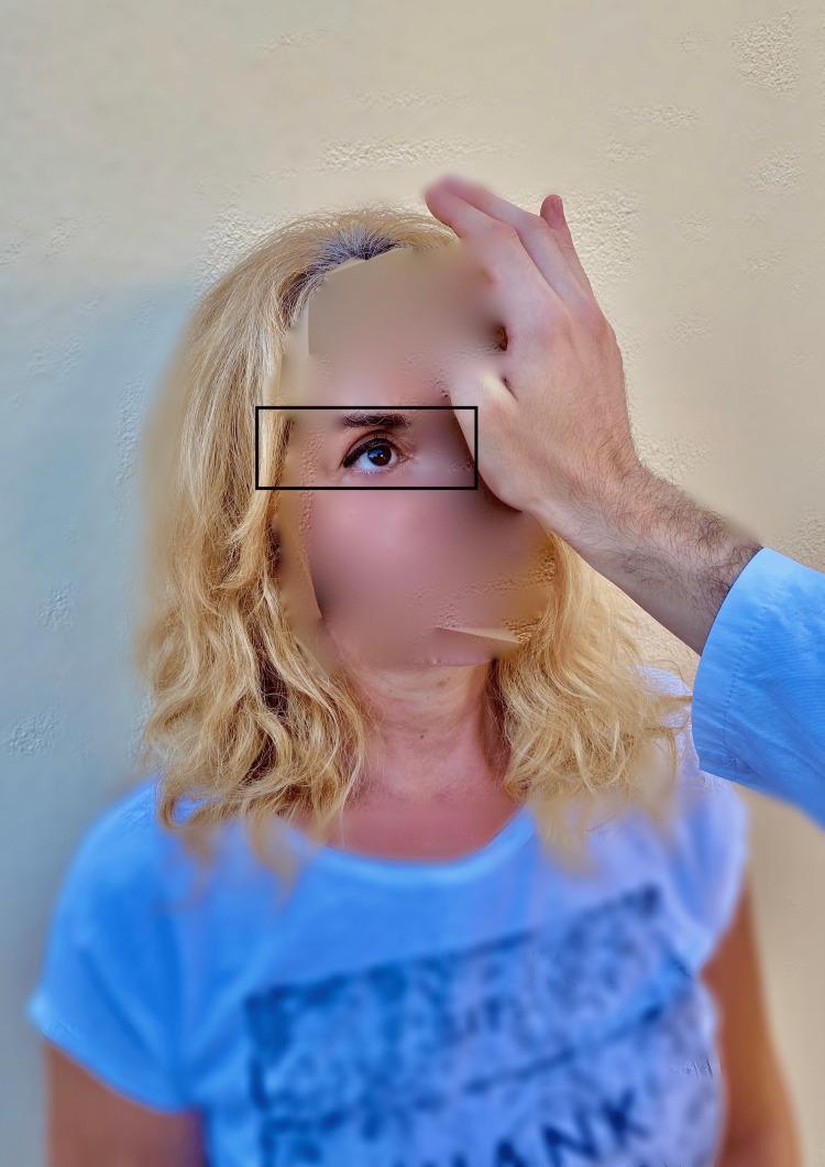
A clinical photograph demonstrating the 'test of skew,' a component of the HINTS (Head Impulse, Nystagmus, Test of Skew) examination used to differentiate central from peripheral causes of acute vestibular syndrome. The image shows an examiner’s hand placed over a patient's left eye while the patient's right eye remains uncovered and is highlighted by a rectangular box to indicate observation. During this maneuver, the examiner performs an alternate cover test while the patient maintains fixation on a central target (typically the examiner’s nose). The clinical focus is to detect skew deviation—a vertical ocular misalignment. In central vestibular disorders, such as a brainstem stroke, uncovering an eye may reveal a corrective vertical shift (refixation saccade). Conversely, the absence of vertical deviation, as seen in this clinical demonstration, is more characteristic of peripheral vestibular conditions like vestibular neuritis. This diagnostic procedure is essential for evaluating patients presenting with continuous vertigo, nystagmus, and nausea.
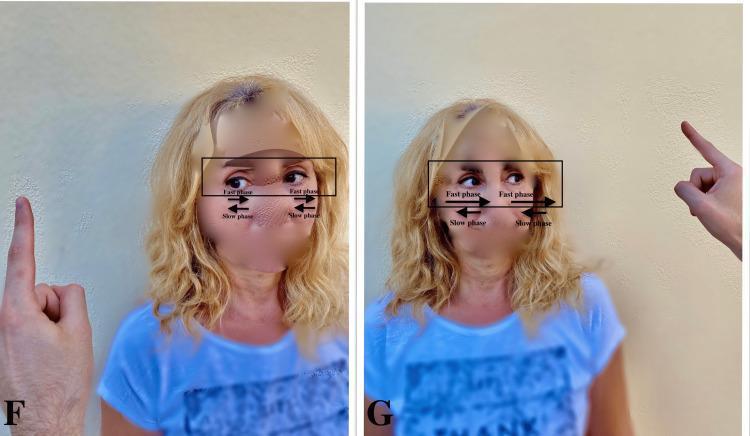
Clinical photograph consisting of two side-by-side images (labeled F and G) demonstrating the assessment of vestibular nystagmus in a patient with acute vestibular loss. In panel F, the examiner's finger is positioned to the patient's left; directional arrows indicate a nystagmus with a slow phase drifting toward the patient's right (the pathologic side) and a fast corrective phase toward the left (the healthy side). In panel G, the examiner's finger moves to the patient's right; annotations illustrate that the nystagmus intensity increases when looking in the direction of the fast phase, consistent with Alexander's Law. This visual educational tool demonstrates the HINTS (Head Impulse, Nystagmus, Test of Skew) examination component used to differentiate peripheral vestibular neuritis from central causes of vertigo, emphasizing that peripheral nystagmus typically has a unidirectional fast phase and follows a specific pattern of slow-phase drift toward the affected ear.
Vertigo - Illustrated Guide
1. The Anatomy Behind Vertigo
- 3 semicircular canals (detect rotational movement - anterior, posterior, lateral)
- Utricle and saccule (detect linear acceleration and gravity)
- Cochlea (for hearing)
2. Classification by Duration - The Big Picture
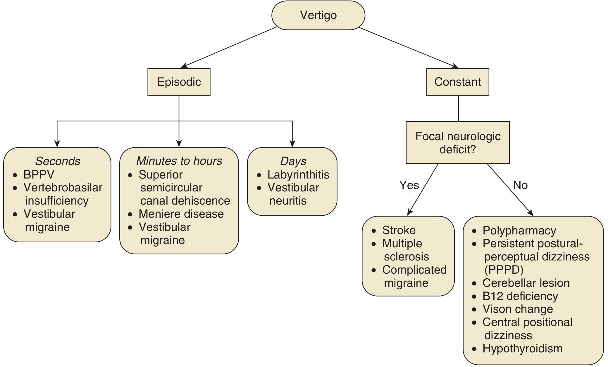
- Seconds of episodic vertigo → BPPV, vertebrobasilar insufficiency, vestibular migraine
- Minutes to hours → Meniere disease, vestibular migraine, superior canal dehiscence
- Days → Labyrinthitis, vestibular neuritis
- Constant + focal neuro deficit → Stroke, MS, complicated migraine (EMERGENCY)
- Constant, no neuro deficit → Drug toxicity (polypharmacy), cerebellar lesion, PPPD, hypothyroidism
3. Most Common Cause: BPPV
How the Epley Maneuver Works
| Step | Position | What happens to otoconia |
|---|---|---|
| 1 | Seated, head rotated 45° toward affected ear | Otoconia sit in posterior canal |
| 2 | Lie back, head extended below table | Debris shifts toward canal apex |
| 3 | Head rotated 90° to the opposite side | Debris moves along canal toward the common crus |
| 4 | Patient rolls onto shoulder (head still rotated) | Debris exits into utricle |
| 5 | Patient sits up | Debris settles in utricle - no longer problematic |
Guideline: Clinicians should treat posterior canal BPPV with canalith repositioning. Routine vestibular suppressant medications (antihistamines, benzodiazepines) should NOT be used for BPPV.
4. Meniere Disease - Endolymphatic Hydrops
Vertigo (hours) + Unilateral tinnitus + Sensorineural hearing loss
5. The HINTS Exam - Ruling Out Stroke
H - Head Impulse Test
- Abnormal (catch-up saccade present) = PERIPHERAL (safe) - the vestibular-ocular reflex is broken peripherally
- Normal = CENTRAL (dangerous) - the brain is compensating, suggesting a central lesion
N - Nystagmus
- Unidirectional horizontal nystagmus = peripheral (vestibular neuritis)
- Direction-changing, vertical, or purely torsional nystagmus = central (stroke)
T - Test of Skew
- Vertical refixation saccade when eye is uncovered = skew deviation = CENTRAL (brainstem lesion)
- No vertical shift = peripheral
HINTS mnemonic for central (dangerous) vertigo: Normal Head impulse + Direction-changing Nystagmus + Skew deviation = CENTRAL STROKE until proven otherwise
6. Peripheral vs. Central Vertigo - Side-by-Side
| Feature | Peripheral | Central |
|---|---|---|
| Common causes | BPPV, vestibular neuritis, Meniere | Stroke, MS, vestibular migraine |
| Onset | Abrupt, intense | Variable |
| Duration | Seconds (BPPV) to days (neuritis) | Constant, weeks |
| Nystagmus | Horizontal/torsional, unidirectional | Vertical, direction-changing |
| Suppressed by fixation? | Yes | No |
| Head impulse test | Abnormal (catch-up saccade) | Normal |
| Skew deviation | Absent | Present |
| Hearing loss | Possible (Meniere, labyrinthitis) | Rare |
| Neurologic signs | Absent | Diplopia, dysarthria, ataxia, weakness |
| Falls | Can walk (unsteady) | Often cannot walk/stand |
| Imaging needed? | Only if atypical or refractory | Urgent MRI |
7. Treatment Summary
| Cause | First-Line Treatment |
|---|---|
| BPPV (posterior canal) | Epley maneuver |
| BPPV (horizontal canal) | Barbecue roll / Gufoni maneuver |
| Vestibular neuritis | Corticosteroids + vestibular rehab |
| Meniere disease | Low-Na diet + diuretics; intratympanic gentamicin for refractory |
| Vestibular migraine | Migraine prophylaxis (topiramate, beta-blockers) |
| Acute symptomatic relief | IV ondansetron 4 mg (1st line in ED) |
| Central/stroke | Urgent neurology, thrombolytics if eligible |
Mujhe ek mbbs MD ko explain Krna hai Isliye mujhe acche se explain kro easy language me
vestibular system balance pathway brainstem cerebellum diagram
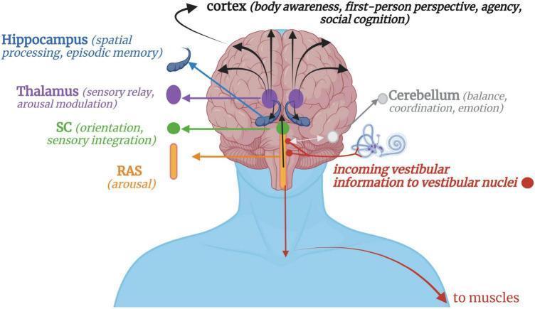
This medical schematic illustrates the neuroanatomical pathways of vestibular processing in the human brain. The diagram depicts the flow of afferent information starting from the inner ear's vestibular apparatus (semicircular canals and otoliths) to the vestibular nuclei in the brainstem. From these nuclei, several projections are shown: a descending pathway to muscles for motor and postural control; a bidirectional connection to the cerebellum for balance, coordination, and emotional processing; and ascending pathways. The ascending signals pass through the reticular activating system (RAS) for arousal modulation and the superior colliculi (SC) for orientation and sensory integration. Information further travels through the thalamus, acting as a sensory relay, to reach the hippocampus (spatial processing and episodic memory) and the cerebral cortex. The cortical destination is linked to higher-order functions including body awareness, first-person perspective, sense of agency, and social cognition. The illustration uses color-coded labels and arrows to represent the complex integration of vestibular signals within the central nervous system.
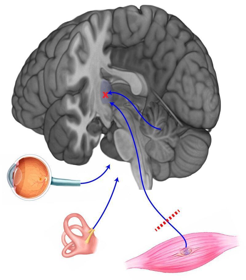
This pathophysiology diagram illustrates the integration of sensory inputs required for balance and postural control. The central feature is a sagittal section of the human brain, highlighting the cerebellum, brainstem, and cerebral cortex. Three primary peripheral systems are shown providing input to the central nervous system: the visual system (represented by a cross-section of the eye), the vestibular system (represented by the inner ear and semicircular canals), and the proprioceptive system (represented by a skeletal muscle with a muscle spindle). Blue arrows trace the afferent pathways from these organs toward a central integration area in the brain. A red 'X' and dashed red lines over the proprioceptive pathway from the muscle indicate a site of dysfunction or sensory interruption. The diagram serves as an educational tool to demonstrate how multisensory integration—combining visual, vestibular, and proprioceptive data—is essential for maintaining equilibrium, and how disruptions in these pathways contribute to clinical balance disorders.
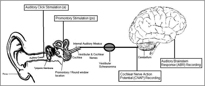
A simplified medical schematic diagram of the human auditory pathway, illustrating key anatomical structures and neurophysiological monitoring sites relevant to vestibular schwannoma surgery. The diagram maps the pathway from the external ear (pinna and auditory canal) to the brain. Labeled structures include the tympanic membrane, promontory/round window location, cochlea, internal auditory meatus, and the vestibular and cochlear nerves. A vestibular schwannoma (acoustic neuroma) is depicted as a mass compressing the auditory nerve. The central nervous system components shown include the cerebellum and the brain. The diagram highlights two stimulation points: 'Auditory Click Stimulation' (acoustic) and 'Promontory Stimulation' (electrical). It also identifies two recording sites for intraoperative neuromonitoring: 'Cochlear Nerve Action Potential (CNAP) Recording' at the level of the cochlear nerve and 'Auditory Brainstem Response (ABR) Recording' at the brainstem level. This illustration serves as an educational guide for understanding the logic behind different auditory monitoring techniques used to preserve hearing during neurosurgical procedures.
nystagmus peripheral central vertigo comparison clinical features
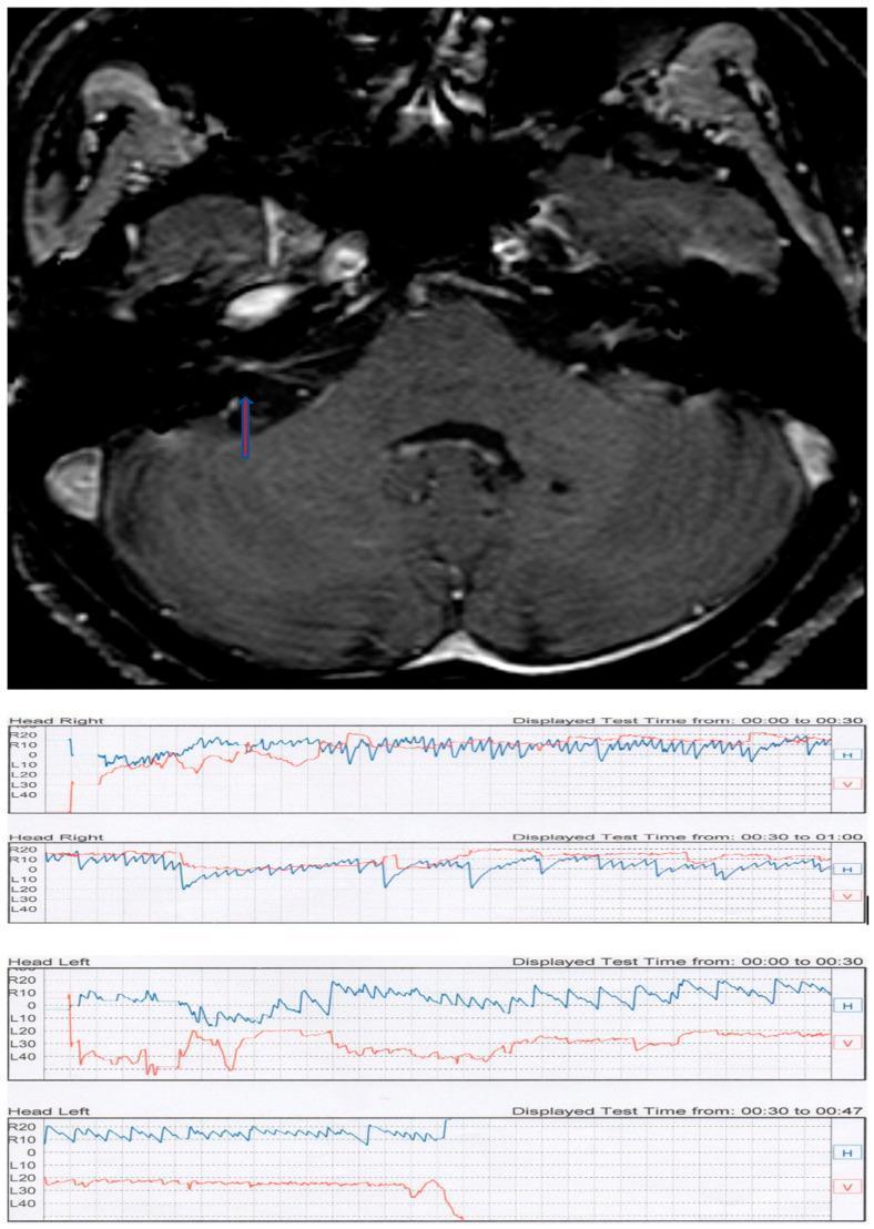
This composite educational figure illustrates the diagnostic findings for a patient with positional vertigo. The top panel features an axial T1-weighted contrast-enhanced MRI scan of the posterior fossa. A blue arrow highlights a small, enhancing lesion within the right internal auditory canal, diagnostic of an intrameatal acoustic neuroma (vestibular schwannoma). The bottom panel contains multiple videonystagmography (VNG) tracings from a Pagnini-McClure positional test. The recordings show horizontal (H, blue line) and vertical (V, red line) eye movements during 'Head Right' and 'Head Left' maneuvers. The tracings demonstrate apogeotropic direction-changing positional nystagmus characterized by rapid, paroxysmal horizontal oscillations. The VNG evidence of central-type positional nystagmus combined with MRI findings demonstrates how a peripheral nerve tumor can manifest with atypical positional vertigo, mimicking or complicating Benign Paroxysmal Positional Vertigo (BPPV) clinical presentations.
A clinical photograph demonstrating the 'test of skew,' a component of the HINTS (Head Impulse, Nystagmus, Test of Skew) examination used to differentiate central from peripheral causes of acute vestibular syndrome. The image shows an examiner’s hand placed over a patient's left eye while the patient's right eye remains uncovered and is highlighted by a rectangular box to indicate observation. During this maneuver, the examiner performs an alternate cover test while the patient maintains fixation on a central target (typically the examiner’s nose). The clinical focus is to detect skew deviation—a vertical ocular misalignment. In central vestibular disorders, such as a brainstem stroke, uncovering an eye may reveal a corrective vertical shift (refixation saccade). Conversely, the absence of vertical deviation, as seen in this clinical demonstration, is more characteristic of peripheral vestibular conditions like vestibular neuritis. This diagnostic procedure is essential for evaluating patients presenting with continuous vertigo, nystagmus, and nausea.
vestibular neuritis labyrinthitis clinical presentation treatment
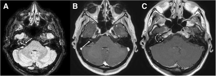
This composite diagnostic image features three axial MRI sequences of the head at the level of the cerebellopontine angle, illustrating vestibular neuritis. Image A is a FLAIR sequence showing mild hyperintensity in the cisternal tract of the right vestibular nerve (white arrow) and the right labyrinth (dotted circle). Images B and C are T1-weighted post-gadolinium sequences. Image B demonstrates pathological linear enhancement of the right vestibular nerve (white arrow) as it traverses towards the internal auditory canal. Image C highlights abnormal contrast enhancement within the right labyrinth (dotted circle). These findings are asymmetric, with no corresponding hyperintensity or enhancement on the contralateral (left) side. Such features indicate a disruption of the blood-nerve barrier and increased vascularity characteristic of inflammatory processes affecting the eighth cranial nerve and inner ear structures. The imaging is critical for differentiating between vestibular neuritis and other pathology like vestibular schwannoma or labyrinthitis.
A series of five intraoral clinical photographs documenting the restorative treatment of a right mandibular second premolar (tooth 45). Image A displays the initial presentation from a vestibular (buccal) view, revealing a significant loss of tooth structure involving the disto-vestibular and occlusal walls, consistent with a failed restoration or extensive caries. The adjacent gingiva shows localized hyperemia and inflammation. Images B (vestibular) and C (occlusal) show the immediate postoperative result of the restoration using aesthetic micro-hybrid composite resin. The restoration successfully anatomically reconstructs the cusps and marginal ridges with a high-luster polish. Images D (vestibular) and E (occlusal) provide a one-and-a-half-year follow-up, demonstrating the clinical longevity of the restoration. In this long-term view, the composite material shows slight color maturation and minor surface wear, yet maintains good marginal integrity and anatomical form. An implant-supported crown is also visible in the adjacent tooth 46 position in the follow-up images.
🌀 VERTIGO - MBBS/MD ke liye Complete Guide
🧠 Pehle Samjho: Balance kaise hota hai?
- Inner ear (Vestibular system) - sab se important
- Eyes (Visual system) - dekh ke balance karo
- Proprioception (Muscles/joints) - body position feel karo
Simple definition: Vertigo = False sensation of motion (usually spinning) - patient ko lagta hai woh ghoom raha hai ya sab kuch ghoom raha hai, jabke actually kuch nahi ghoom raha.
🌐 Vestibular Pathway - Signal kahan jaata hai?
- Cerebellum - coordination aur balance
- Thalamus - sensory relay
- Cortex - conscious body awareness
- Muscles - postural control (Vestibulospinal tract)
- Eyes - VOR (Vestibulo-Ocular Reflex) - isi se nystagmus hoti hai
🗺️ Diagnosis ka Framework - Duration se socho
| Duration | Socho kya ho sakta hai |
|---|---|
| Seconds (position se trigger) | BPPV |
| Minutes to Hours | Meniere disease, Vestibular migraine |
| Days | Vestibular neuritis, Labyrinthitis |
| Constant + Neuro signs | 🚨 Stroke, MS, Tumor |
📦 Causes - Peripheral vs Central
⚪ PERIPHERAL CAUSES (inner ear / 8th nerve)
1. BPPV - Benign Paroxysmal Positional Vertigo
Sabse common cause - approx 40% cases
- Brief spinning (<30 seconds)
- Position change se trigger (seedha uthna, neecha dekhna, lait jaana)
- Nausea/vomiting ho sakti hai
- Hearing loss NAHI hoti
2. Vestibular Neuritis / Labyrinthitis
- Neuritis = sirf vestibular branch affect → hearing normal
- Labyrinthitis = cochlea bhi affect → hearing loss bhi hogi
- Sudden onset severe vertigo (hours se days)
- Nausea/vomiting bahut zyada
- Spontaneous nystagmus - lesion ki OPPOSITE side mein fast phase
- Head impulse test ABNORMAL (catch-up saccade)
- Viral URTI pehle ho sakta hai
Panel A (FLAIR): Right vestibular nerve aur labyrinth mein hyperintensity (white arrow + dotted circle). Panel B & C (T1 + Gad): Enhancement of right vestibular nerve - blood-nerve barrier disruption from inflammation.
- Methylprednisolone - recovery speed karta hai
- Vestibular suppressants for acute phase (meclizine, ondansetron)
- Vestibular rehabilitation jab acute phase khatam ho
3. Meniere Disease
🔊 Hearing loss (sensorineural, unilateral, fluctuating) + 🎵 Tinnitus (low-frequency, roaring) + 🌀 Vertigo (hours)
- Episodes last 20 min - 24 hours
- Ear fullness/pressure feel hoti hai before episode (aura)
- Initially low-frequency hearing loss, eventually permanent
- Bilateral ho sakta hai (10-15%)
Left ear mein zyada distension (black arrow). Panel C mein cochlear modiolar area left (2.33 mm²) << right (3.53 mm²) - chronic damage.
- Low sodium diet + diuretics (HCTZ + triamterene)
- Betahistine (controversial, widely used)
- Intratympanic steroids / gentamicin refractory cases mein
🔴 CENTRAL CAUSES (Brainstem / Cerebellum)
Dangerous - inka miss karna fatal ho sakta hai
| Cause | Clue |
|---|---|
| Posterior circulation stroke | Sudden onset + vascular risk factors |
| Cerebellar hemorrhage | Severe headache + ataxia + cannot walk |
| Wallenberg syndrome (PICA) | Crossed sensory loss + Horner syndrome |
| Vestibular migraine | Young female + headache history |
| Multiple sclerosis | Young adult + multiple episodes |
🔍 HINTS Exam - Stroke ko BPPV se kaise alag karo?
HINTS = Head Impulse + Nystagmus + Test of Skew
H - Head Impulse Test
| Result | Meaning |
|---|---|
| Abnormal (catch-up saccade) | 👍 Peripheral (VOR broken) - Safe |
| Normal | 🚨 Central (VOR intact) - Stroke suspect |
N - Nystagmus
| Pattern | Meaning |
|---|---|
| Unidirectional horizontal | Peripheral (vestibular neuritis) |
| Direction-changing / Vertical | 🚨 Central |
T - Test of Skew
- Vertical correction saccade aaye → Skew deviation → 🚨 Central (brainstem lesion)
- Koi correction nahi → Peripheral
HINTS mnemonic for CENTRAL (dangerous): Normal head impulse + Inappropriate (direction-changing) nystagmus + Test of Skew positive = STROKE jab tak prove nahi hota
⚡ Peripheral vs Central - Side-by-Side Table
| Feature | 🟢 Peripheral | 🔴 Central |
|---|---|---|
| Onset | Abrupt, intense | Variable |
| Course | Episodic / days | Constant / progressive |
| Nystagmus direction | Horizontal/torsional - unidirectional | Vertical ya direction-changing |
| Nystagmus suppressed by fixation | Haan | Nahi |
| Head Impulse Test | Abnormal (saccade present) | Normal |
| Skew deviation | Absent | Present |
| Hearing loss | Possible (Meniere, labyrinthitis) | Rare |
| Neurological signs | Absent | Present - dysarthria, diplopia, ataxia |
| Walk kar sakta hai? | Unsteady but walks | Often cannot stand/walk |
| Imaging | MRI only if atypical | Urgent MRI |
💊 Treatment Summary
| Cause | Treatment |
|---|---|
| BPPV | ✅ Epley maneuver (1st line, highly effective) |
| Vestibular neuritis | Methylprednisolone + vestibular rehab |
| Meniere disease | Low Na + diuretics + betahistine |
| Acute symptomatic relief | IV Ondansetron 4mg (1st choice ED mein) |
| Severe nausea/vomiting | IM Promethazine (IV avoid - FDA warning) |
| Posterior fossa stroke | Urgent neurology - thrombolytics if eligible |
| Vestibular migraine | Migraine prophylaxis - topiramate, beta-blockers |
Yaad rakho: Meclizine/antihistamines BPPV mein routinely mat do (guideline recommendation against). Ye central compensation ko slow karte hain. Sirf acute severe nausea ke liye use karo.
🚨 Red Flags - Kabhi mat chuko
- 🔴 Khada nahi ho sakta / walk nahi kar sakta
- 🔴 Vertical ya direction-changing nystagmus
- 🔴 Normal head impulse test (despite constant vertigo)
- 🔴 Sudden severe headache saath
- 🔴 Diplopia, dysarthria, dysphagia
- 🔴 Crossed sensory loss (Wallenberg)
- 🔴 Horner syndrome
- 🔴 Skew deviation positive
- 🔴 Age >60 + hypertension/DM/AF + new onset isolated vertigo
📝 Quick Revision Box (Exam ke liye)
BPPV     → Seconds + Positional + Dix-Hallpike + Epley
Neuritis → Days + Sudden + No hearing loss + Abnormal HIT
Meniere  → Hours + Triad (HVT) + Endolymphatic hydrops
Stroke   → Constant + Neuro signs + Normal HIT = DANGER

HINTS = Central hoga agar:
Normal HIT + Direction-changing nystagmus + Skew +ve