
Hydrocele
I'll start by reading the medical library skill, then search for comprehensive information on hydrocele.
Reading File
Now let me search the medical library for hydrocele content in parallel:
Finding Sources
Searching Images
hydrocele scrotal swelling transillumination
Excellent - multiple sources found. Let me read the most comprehensive ones in parallel:
Reading File
Reading File
Reading File
Bailey & Love has the best content. Let me read more from it and also fetch the diagram:
Reading File
Loading Image
Reading File
Let me get the remaining treatment content and the Campbell-Walsh content on hydrocele in pediatric urology:
Reading File
Finding Sources
Let me fetch the clinical photo and also read the filarial hydrocele content:
Loading Image
Reading File
I now have comprehensive information to compose a thorough answer. Let me also fetch the surgical procedure images:
Loading Image
I now have all the information needed for a comprehensive answer. Here is a complete, well-structured overview of hydrocele:
Hydrocele
A hydrocele is an abnormal collection of serous fluid in a part of the processus vaginalis - usually the tunica vaginalis surrounding the testis, and occasionally along the spermatic cord.
Epidemiology
- Affects an estimated 1% of adult men
- Over 80% of newborn boys have a patent processus vaginalis, but most close spontaneously within 18 months of age
- Secondary hydroceles usually occur in men >40 years
(Bailey and Love's Short Practice of Surgery 28th Edition)
Embryology & Pathophysiology
The processus vaginalis is a diverticulum of the peritoneal cavity. It descends with the testes into the scrotum via the inguinal canal around the 28th week of gestation, with gradual closure through infancy and childhood.
Classification (Types)
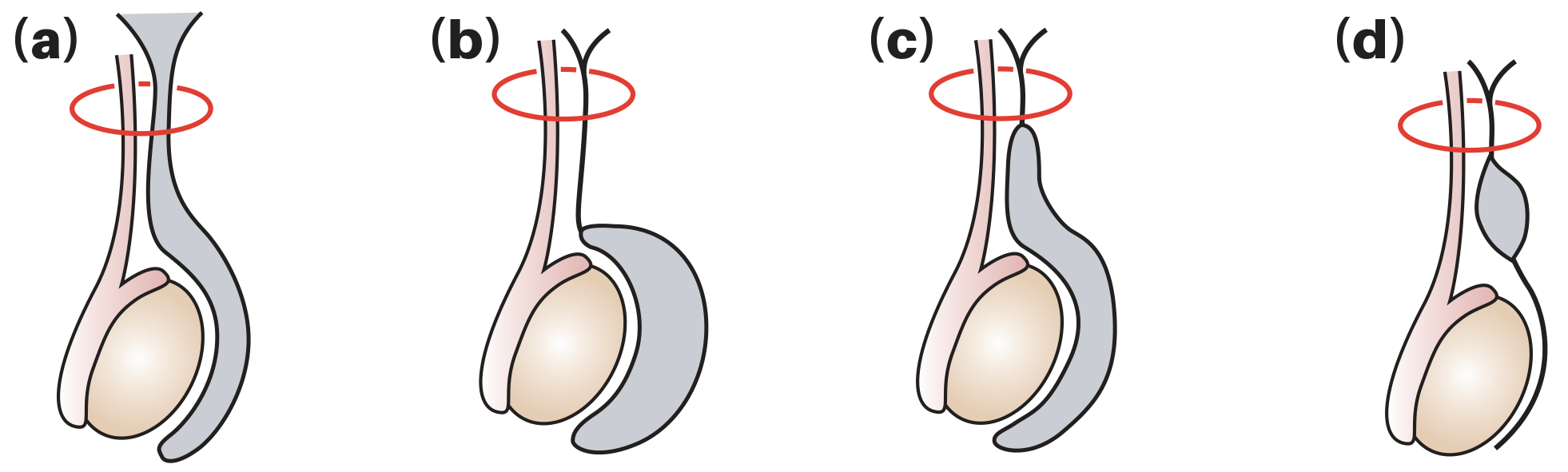
| Type | Mechanism |
|---|---|
| 1. Communicating (Congenital) | Patent processus vaginalis allows peritoneal fluid to flow freely into the scrotum. Often associated with indirect inguinal hernias. |
| 2. Non-communicating (Vaginal) | Processus vaginalis closed; fluid accumulates due to excessive production or defective absorption by tunica vaginalis (disrupted lymphatic drainage). The most common type globally. If idiopathic = "primary" hydrocele. |
| 3. Infantile hydrocele | Distal end closes correctly; mid-portion remains patent with proximal end open - communicates with tunica vaginalis. |
| 4. Hydrocele of the cord | Distal and proximal ends both closed; mid-portion remains patent - isolated cystic collection along the cord. |
| 5. Abdominoscrotal hydrocele | Uncommon (1.25% of all hydroceles); non-communicating tense scrotal mass extending into the abdomen; ~30% bilateral. |
(Bailey and Love's 28th Ed; Campbell-Walsh-Wein Urology)
Secondary Causes
Secondary hydroceles result from local pathology:
- Infection / epididymo-orchitis
- Testicular torsion
- Testicular neoplasm (important to exclude - do not puncture if tumour suspected due to risk of malignant needle-track implantation)
- Trauma
- Radiotherapy
- Filariasis (Wuchereria bancrofti - accounts for up to 80% of hydroceles in tropical countries)
- Post-varicocelectomy (due to lymphatic obstruction; incidence 3-33%, average ~7%)
Clinical Features
Examination (upright and supine)
Ask three key questions:
- Can you get above the swelling? - If no, consider inguinal hernia entering the scrotum.
- Is the testis/epididymis palpable? - A hydrocele encloses both structures so they may be impalpable, but you can get "above" the swelling to palpate a normal spermatic cord.
- Does it transilluminate? - Hydroceles are typically translucent and transilluminate brightly.
Presentation
- Usually painless - may reach significant size before presentation
- Congenital hydrocele may be intermittent (fluid drains into peritoneum when lying down)
- Be wary of an acute hydrocele in a young man - may be a testicular tumour
- Hydrocele of the cord: smooth oval swelling above testis near spermatic cord; moves downwards and becomes less mobile when testis is pulled downwards
- Hydrocele of the canal of Nuck: female equivalent; cyst along round ligament, partially within inguinal canal
- If bilateral congenital hydroceles - check for ascites
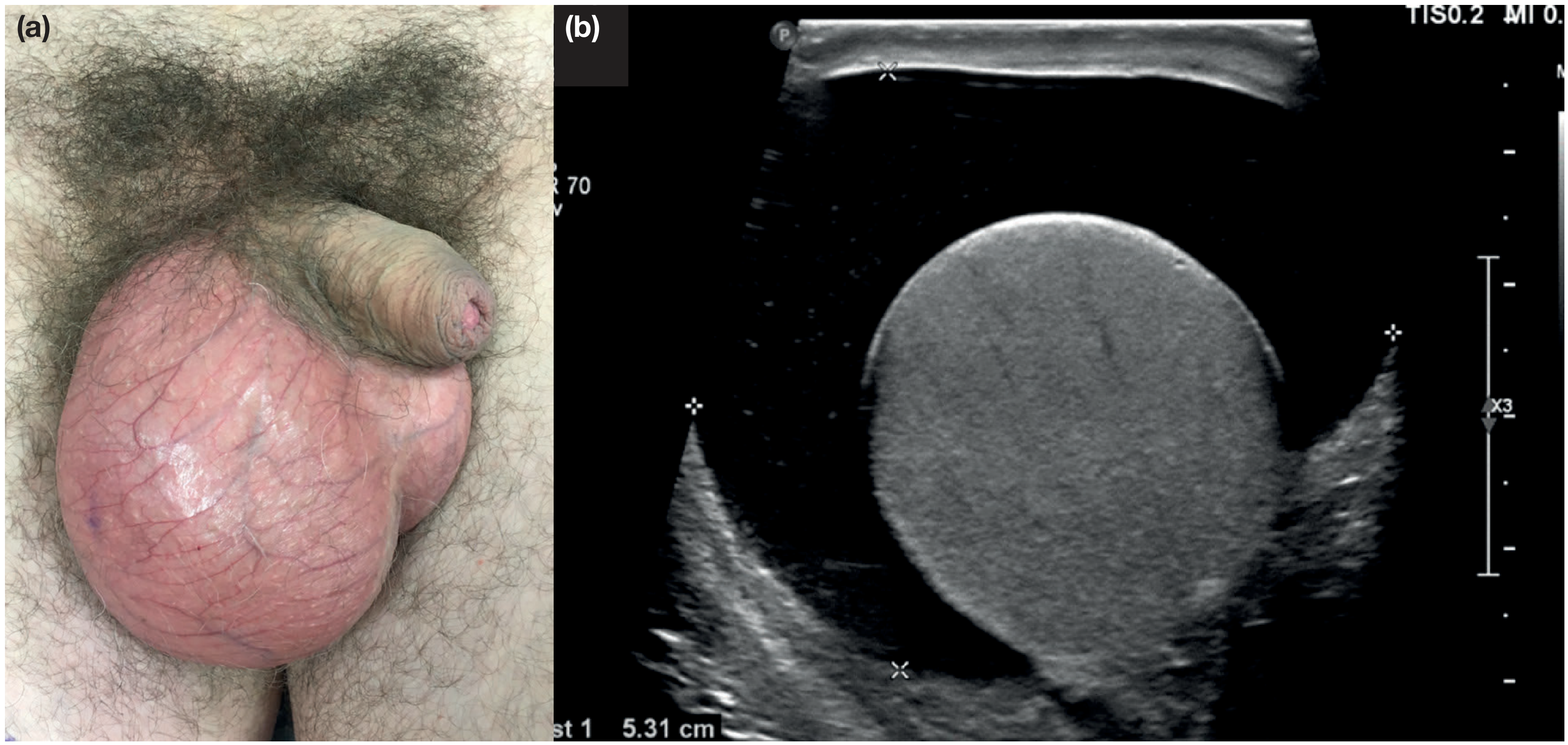
Clinical Image & Ultrasound
Investigations
- Scrotal ultrasound - valuable adjunct in nearly all cases; assesses the testis itself for tumour or other pathology, especially when testis/epididymis are impalpable
- Tumour markers (AFP, beta-hCG, LDH) if malignancy suspected
Treatment
1. Conservative
- Small, asymptomatic hydroceles do not require treatment
- Congenital hydroceles frequently resolve spontaneously by 18 months
2. Surgical (main treatment)
Three established techniques:
A. Lord's Operation (Plication)
- Suitable for thin-walled sacs
- Interrupted absorbable sutures plicate the redundant tunica vaginalis, bunching it at its testicular attachment
- Minimal dissection; reduced risk of haematoma
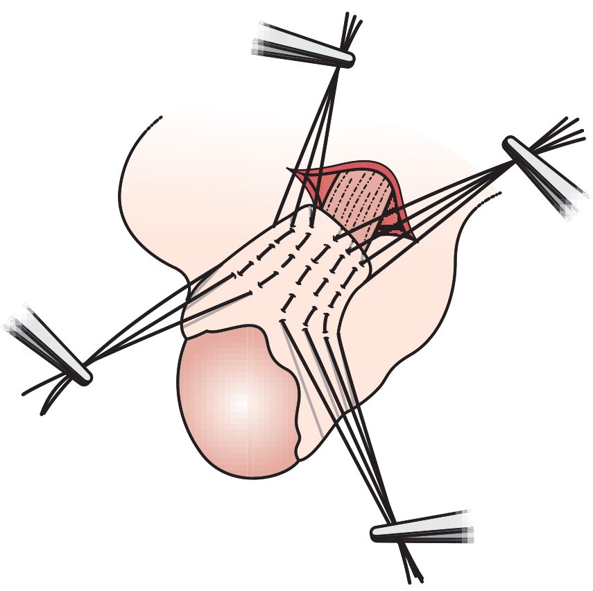
B. Jaboulay's Procedure (Eversion)
- Hydrocele sac is opened and everted behind the testis, then anchored with sutures (testis placed in a fascial pouch)
- Care needed to control bleeding from cut edge to avoid scrotal haematoma
- Overrunning stitches at cut edge reduce this risk
C. Excision
- For thick-walled acquired hydroceles
- Established acquired hydroceles often have thick walls requiring partial or complete sac excision
For congenital/communicating hydrocele: Ligation of the patent processus vaginalis (herniotomy) if no spontaneous resolution.
3. Aspiration ± Sclerotherapy
- Fluid always reaccumulates within a week or so after simple aspiration
- Suitable only for men unfit for surgery
- Risk: bleeding into sac causing haematocele
- Sclerosant injection (e.g. tetracycline, polidocanol) can be effective but is painful; long-term data exist for polidocanol
Special Notes
Post-varicocelectomy Hydrocele
- Average incidence ~7% (range 3-33%) after non-microscopic varicocelectomy
- Caused by lymphatic obstruction
- At least half grow large enough to warrant surgical excision
- Microsurgical technique with magnification to preserve lymphatics almost eliminates this risk
Filarial Hydrocele / Chylocele
- Up to 80% of hydroceles in tropical countries (W. bancrofti endemic areas)
- Follow repeated filarial epididymo-orchitis attacks
- Fluid may contain liquid fat (chyle) from ruptured lymphatic varix - cholesterol-rich
- Dense adhesions in longstanding cases
- Treatment: rest and aspiration; chronic cases require excision of the sac
After Hernia Repair
- If a distal sac or hydrocele is present during inguinal hernia repair, it must be opened widely, not tied off - tying creates a closed space where a new hydrocele can form
Key Points Summary
| Feature | Detail |
|---|---|
| Definition | Fluid in tunica vaginalis |
| Most common sign | Transillumination |
| Investigation of choice | Scrotal ultrasound |
| Contraindication to aspiration | Suspected testicular tumour |
| Mainstay of treatment | Surgery |
| Operation for thin-walled sac | Lord's (plication) |
| Operation for thick-walled sac | Jaboulay's (eversion) or excision |
| Tropical cause | Wuchereria bancrofti (filariasis) |
(Bailey and Love's Short Practice of Surgery 28th Ed, pp. 1586-1587; Campbell-Walsh-Wein Urology)
This is a shared conversation. Sign in to Orris to start your own chat.