
Generalized purpura with thrombocytopenia evaluation, probable diagnosis,other symptoms and signs and management
"immune thrombocytopenia" AND purpura
ASH immune thrombocytopenia guidelines diagnosis management adults children
https://www.hematology.org/education/clinicians/guidelines-a…
clinical photograph generalized petechiae purpura thrombocytopenia
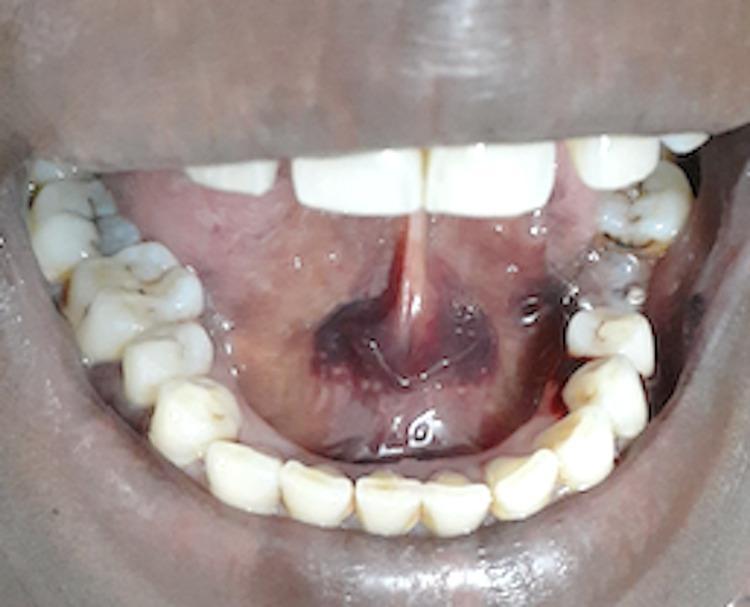
This clinical photograph shows an intraoral view of the floor of the mouth, demonstrating a significant area of submucosal ecchymosis and purpura. A large, irregular, dark purplish-red patch of extravasated blood is visible on the anterior floor of the mouth, bilaterally distributed across the lingual frenum. The lesion is flat (non-raised) and submucosal in nature. Surrounding the primary ecchymosis are smaller, more discrete petechiae and purpuric spots. The adjacent oral mucosa and the gingiva of the mandibular teeth appear otherwise healthy, without obvious swelling or generalized inflammation, though a focal area of possible gingival bleeding is noted in the posterior mandibular quadrant. This visual evidence is highly characteristic of thrombocytopenic purpura, which in this clinical context is associated with phenytoin-induced severe thrombocytopenia. The image serves as a clinical reference for identifying oral manifestations of systemic hematologic disorders and adverse drug reactions.
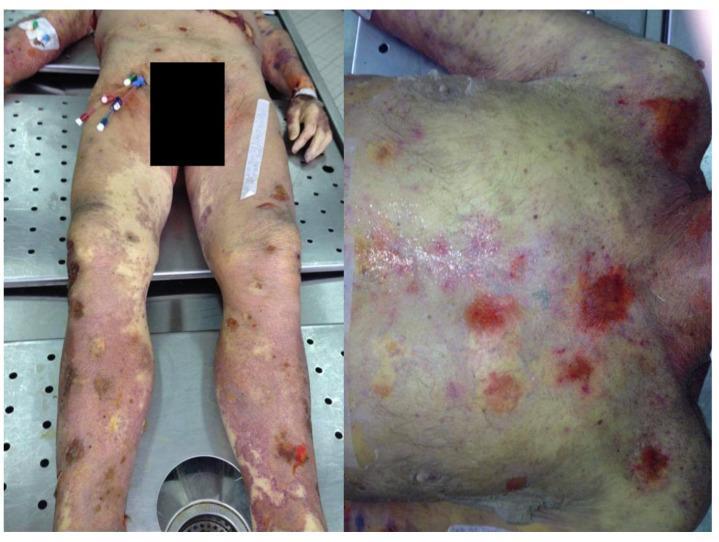
This clinical photograph displays a post-mortem examination of a 71-year-old male, illustrating severe dermatological manifestations of methotrexate toxicity. The left panel shows the lower body and legs, while the right panel provides a close-up of the upper torso. The skin exhibits widespread, generalized erythema and significant jaundice (icterus). There is extensive involvement of both petechiae (pinpoint hemorrhages) and purpura, appearing as irregular, confluent reddish-purple patches across the chest, abdomen, and limbs. Key clinical features include multiple areas of epithelial exfoliation and denuded skin, characteristic of toxic epidermal necrolysis (TEN). Additionally, pre-existing psoriatic plaques are visible on the lower extremities, some appearing eroded or ulcerated. The image serves as a significant educational example of drug-induced severe cutaneous adverse reactions (SCARs) and the systemic effects of bone marrow suppression, such as thrombocytopenia leading to diffuse hemorrhagic lesions.
"immune thrombocytopenia" AND treatment
Generalized purpura with thrombocytopenia
Probable diagnosis
History and examination
Symptoms and signs of thrombocytopenic purpura
- Pinpoint, non-blanching petechiae, often initially on dependent areas such as the legs
- Generalized purpura and easy bruising/ecchymoses
- Epistaxis, gingival bleeding
- Oral blood blisters or “wet purpura”
- Menorrhagia or prolonged bleeding after minor trauma
- Hematuria, melena, hematemesis in severe disease
- Headache, vomiting, altered consciousness, or focal neurology may indicate intracranial bleeding
Look specifically for
| Finding | Diagnostic implication |
|---|---|
| Isolated petechiae/purpura, otherwise well | ITP or drug-induced thrombocytopenia |
| Fever, confusion, seizures, renal impairment | TTP, sepsis, or other thrombotic microangiopathy |
| Jaundice, pallor, dark urine | Hemolysis, including TTP |
| Hepatosplenomegaly or lymphadenopathy | Leukemia/lymphoma, liver disease, hypersplenism, infection |
| Bone pain, weight loss, recurrent infections | Marrow infiltration/failure or hematologic malignancy |
| Recent diarrhea | Shiga toxin-associated HUS |
| Recent heparin exposure plus thrombosis | Heparin-induced thrombocytopenia |
| New medicine, herbal, OTC drug, quinine-containing product | Drug-induced thrombocytopenia |
| Malarial exposure, fever, travel | Infection-related thrombocytopenia |
| Rash plus arthralgia, photosensitivity, renal symptoms | SLE or vasculitis |
Evaluation
1. Establish urgency
- Active major bleeding or hemodynamic instability
- Suspected intracranial bleed or new neurologic symptoms
- Platelet count usually <10 × 10⁹/L, particularly with bleeding
- Severe mucosal bleeding or wet purpura
- Anemia with schistocytes, renal dysfunction, fever, or neurologic change
- Suspected TTP, DIC, acute leukemia, or sepsis
2. Core investigations
- Repeat CBC with platelet count
- Confirm the result and assess anemia/leukopenia.
- Peripheral blood smear
- Platelet clumps: pseudothrombocytopenia
- Schistocytes: TTP/HUS/DIC or another microangiopathic hemolytic anemia
- Blasts: acute leukemia
- Giant platelets: possible inherited thrombocytopenia or peripheral platelet destruction
- Coagulation screen: PT/INR, aPTT, fibrinogen, D-dimer
- Abnormal coagulation tests and low fibrinogen suggest DIC rather than ITP.
- Hemolysis screen: reticulocyte count, LDH, indirect bilirubin, haptoglobin, direct antiglobulin test
- Renal and liver function tests, urinalysis
- Pregnancy test in people who could be pregnant
- HIV and hepatitis C testing in suspected ITP; consider hepatitis B before immunosuppressive therapy
- Targeted investigations based on context:
- ANA/dsDNA and complement if SLE is suspected
- Malaria testing where relevant
- Blood cultures and infection evaluation if febrile
- ADAMTS13 activity/inhibitor sample if TTP is suspected, but do not delay treatment
- HIT evaluation using a 4Ts assessment and PF4 testing if compatible heparin exposure
3. Bone marrow examination
- Age over 60 years
- Other cell lines are abnormal
- Blasts/dysplasia or atypical cells on smear
- Hepatosplenomegaly/lymphadenopathy
- No response to initial ITP treatment or diagnosis remains unclear
Key differential diagnoses
1. Immune thrombocytopenia
- Isolated thrombocytopenia
- Normal coagulation profile
- No schistocytes or systemic illness
- Petechiae, bruising, mucosal bleeding
- Primary or secondary to HIV, hepatitis C, SLE, etc.
2. TTP: do not miss
3. DIC
- Often sepsis, trauma, obstetric catastrophe, malignancy
- Thrombocytopenia plus prolonged PT/aPTT, high D-dimer, falling fibrinogen, and schistocytes
4. Drug-induced thrombocytopenia
5. Bone-marrow disorders
- Acute leukemia
- Aplastic anemia
- Myelodysplastic syndrome
- Marrow infiltration
- Usually associated with anemia and/or leukopenia, abnormal smear, constitutional symptoms, or organomegaly.
Management
General measures
- Admit if significant bleeding, very low platelet count, diagnostic uncertainty, or concern for TTP/DIC/sepsis.
- Avoid IM injections, rectal procedures, contact sports, aspirin, NSAIDs, and unnecessary antiplatelet/anticoagulant drugs.
- Treat any underlying trigger: infection, secondary autoimmune disease, liver disease, or drug exposure.
- Use local measures and antifibrinolytic treatment when appropriate for mucosal bleeding, under clinician supervision.
If likely ITP without major bleeding
- Observation may be appropriate for no/minor bleeding and a safe platelet count.
- Short-course corticosteroids are conventional first-line therapy for patients needing treatment.
- Prednisone or high-dose dexamethasone are commonly used.
- Avoid prolonged corticosteroid courses if possible because of toxicity.
- IVIG is useful where a rapid platelet rise is needed, such as clinically important bleeding, very low count, or urgent procedure.
- Anti-D immunoglobulin may be an option in selected Rh-positive, non-splenectomized patients.
- For persistent/chronic disease, hematology-directed options include:
- Thrombopoietin receptor agonists: eltrombopag, romiplostim, avatrombopag
- Rituximab
- Fostamatinib in selected refractory cases
- Splenectomy, generally deferred until persistent disease and after appropriate vaccination/counseling
If major or critical bleeding in ITP
- IV corticosteroids
- IVIG
- Platelet transfusion for life-threatening or significant active bleeding, generally alongside IVIG and steroids because transfused platelets are otherwise rapidly destroyed
- Site-specific management, for example neuroimaging and neurosurgical input if intracranial hemorrhage is possible
If TTP is suspected
- Draw ADAMTS13 samples if feasible, but start treatment immediately
- Urgent daily therapeutic plasma exchange with plasma replacement
- Corticosteroids
- Specialist-directed caplacizumab and immunosuppression such as rituximab when appropriate
- Do not rely on platelet transfusion unless bleeding is life-threatening, because it can worsen thrombosis
If DIC or sepsis is suspected
- Treat the underlying cause urgently, especially sepsis or obstetric/traumatic triggers.
- Use blood products based on bleeding, procedures, and coagulation results, not platelet count alone.