
Neck dissection types and complications, classify lymphnode of neck with boundaries along with images, both these answers in detail according to standard ENT textbook and in simplified manner
Reading File
Quick Answer: Neck dissections are classified as Radical, Modified Radical (Types I-III), Selective, and Extended. The neck lymph nodes are divided into 6 levels (I-VI) by the AAO-HNS classification. Complications include nerve injuries (XI, XII, phrenic), vascular injuries, chyle leak, and lymphedema.
Searching sources...
Finding Sources
Finding Sources
Finding Sources
Reading File
Reading File
Finding Sources
Reading File
Finding Sources
Reading File
Reading File
Excellent data retrieved. Now let me get the complications and the Scott-Brown classification table, plus search for images.
Finding Sources
Reading File
Searching Images
neck dissection radical modified selective anatomy
Reading File
Searching Images
cervical lymph node levels I II III IV V VI neck anatomy illustration
Reading File
Searching Images
spinal accessory nerve injury shoulder drop after neck dissection
Reading File
Searching Images
neck lymph node zones levels submental submandibular jugular posterior triangle
Reading File
I have comprehensive data. Let me now check for images in the textbook and get the Scott-Brown classification table image.
Finding Sources
Reading File
Reading File
I now have all the key data including the image URLs. Let me compile the comprehensive answer.---
PART 1 - Classification of Cervical Lymph Nodes (AAO-HNS/Memorial Sloan Kettering Level System)
Source: Cummings Otolaryngology Head and Neck Surgery; Scott-Brown's Otorhinolaryngology Head & Neck Surgery; Fischer's Mastery of Surgery 8e
The neck lymph nodes are grouped into 6 main levels (I-VI), with Level VII added by some authorities for the superior mediastinum. This system was originally described by the Memorial Sloan Kettering Group and is now standardized by the American Academy of Otolaryngology.
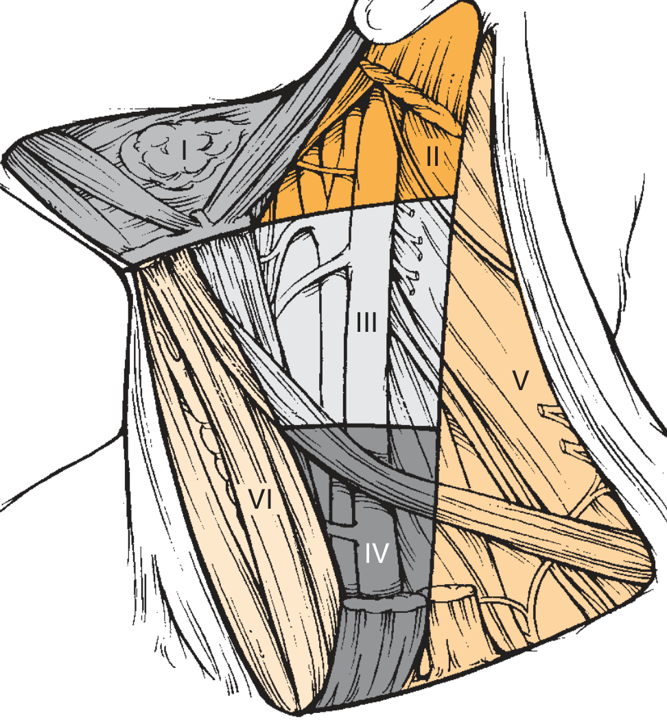
Fig. 118.1 - The six levels of the neck (Cummings Otolaryngology). Level I: submental and submandibular; Level II: upper jugular; Level III: middle jugular; Level IV: lower jugular; Level V: posterior triangle; Level VI: anterior compartment.
LEVEL I - Submental and Submandibular Group
Level Ia - Submental Triangle
| Boundary | Structure |
|---|---|
| Superior | Symphysis of mandible |
| Inferior | Hyoid bone |
| Anterior (medial) | Left anterior belly of digastric |
| Posterior (lateral) | Right anterior belly of digastric |
Clinical note: Nodes lie within the submental triangle, bounded by the anterior bellies of both digastric muscles and the hyoid bone.
Level Ib - Submandibular Triangle
| Boundary | Structure |
|---|---|
| Superior | Body of mandible |
| Inferior | Posterior belly of digastric |
| Anterior (medial) | Anterior belly of digastric |
| Posterior (lateral) | Stylohyoid muscle |
Clinical note: The submandibular gland (SG) is routinely removed during dissection here to ensure complete nodal clearance. Perifacial nodes (Nodes of Stahr/buccinator nodes) lie outside this triangle and must be included for lip/buccal/cheek primaries.
Drains from: Oral cavity (floor of mouth, anterior tongue, lower lip, chin, anterior hard palate), cheek, nasal vestibule.
LEVEL II - Upper Jugular Group
Located around the upper third of the internal jugular vein (IJV) from the skull base to the carotid bifurcation (or hyoid bone clinically). Divided by the spinal accessory nerve (SAN) into IIa and IIb.
Level IIa - Anterior to SAN
| Boundary | Structure |
|---|---|
| Superior | Lower bony margin of jugular fossa (skull base) |
| Inferior | Level of lower body of hyoid bone |
| Anterior (medial) | Stylohyoid muscle |
| Posterior (lateral) | Vertical plane defined by spinal accessory nerve |
Level IIb - Posterior to SAN ("Submuscular Recess")
| Boundary | Structure |
|---|---|
| Superior | Lower bony margin of jugular fossa |
| Inferior | Level of lower body of hyoid bone |
| Anterior (medial) | Vertical plane defined by SAN |
| Posterior (lateral) | Posterior border of SCM |
Key point: IIb nodes lie in the "submuscular recess" posterolateral to the SAN. Routine dissection of IIb may be omitted in clinically N0 laryngeal cancer as metastatic risk is low.
Drains from: Oral cavity, nasal cavity, nasopharynx, oropharynx, hypopharynx, larynx, parotid gland.
LEVEL III - Middle Jugular Group
| Boundary | Structure |
|---|---|
| Superior | Level of lower body of hyoid bone |
| Inferior | Horizontal plane along inferior border of anterior cricoid arch |
| Anterior (medial) | Lateral border of sternohyoid muscle |
| Posterior (lateral) | Posterior border of SCM (or sensory branches of cervical plexus) |
Memory trick: Level III = from hyoid to cricoid.
Drains from: Oral cavity, nasopharynx, oropharynx, hypopharynx, larynx, thyroid.
LEVEL IV - Lower Jugular Group
| Boundary | Structure |
|---|---|
| Superior | Horizontal plane along inferior border of anterior cricoid arch |
| Inferior | Clavicle |
| Anterior (medial) | Lateral border of sternohyoid muscle |
| Posterior (lateral) | Posterior border of SCM (or sensory branches of cervical plexus) |
Memory trick: Level IV = from cricoid to clavicle.
Drains from: Hypopharynx, larynx, thyroid, esophagus, trachea. Skip metastases from infraclavicular primaries (stomach, lung, breast) can occur here - the classic "Virchow's node" (left Level IV/supraclavicular).
LEVEL V - Posterior Triangle Group
Divided by a horizontal plane at the level of the inferior border of the cricoid arch:
Level Va - Upper Posterior Triangle (Spinal Accessory nodes)
| Boundary | Structure |
|---|---|
| Superior | Convergence of SCM and trapezius muscles (apex of posterior triangle) |
| Inferior | Horizontal plane at inferior border of anterior cricoid arch |
| Anterior (medial) | Posterior border of SCM |
| Posterior (lateral) | Anterior border of trapezius muscle |
Level Vb - Lower Posterior Triangle (Transverse cervical and supraclavicular nodes)
| Boundary | Structure |
|---|---|
| Superior | Horizontal plane at inferior border of anterior cricoid arch |
| Inferior | Clavicle |
| Anterior (medial) | Posterior border of SCM |
| Posterior (lateral) | Anterior border of trapezius muscle |
Drains from: Nasopharynx, oropharynx, posterior scalp, posterior neck skin. Nasopharyngeal carcinoma classically metastasizes to Level V.
LEVEL VI - Anterior Compartment Group (Central Compartment)
| Boundary | Structure |
|---|---|
| Superior | Hyoid bone |
| Inferior | Sternal notch |
| Lateral (both sides) | Common carotid artery |
Contains: Pretracheal nodes, precricoid (Delphian node), perithyroidal nodes, paratracheal nodes.
Drains from: Thyroid gland, larynx, subglottis, pyriform sinus apex, trachea, upper esophagus. This level is the primary drainage for thyroid cancers.
LEVEL VII - Superior Mediastinal Group (added by some systems)
| Boundary | Structure |
|---|---|
| Superior | Sternal notch |
| Inferior | Innominate (brachiocephalic) artery |
| Lateral (both sides) | Common carotid artery |
Quick Reference Summary Table
| Level | Name | Key Landmark | Primary Drainage From |
|---|---|---|---|
| Ia | Submental | Anterior bellies of digastric, hyoid | Lower lip, chin, anterior floor of mouth |
| Ib | Submandibular | Body of mandible, digastric | Oral cavity, anterior nasal cavity |
| IIa | Upper jugular (ant) | Skull base to hyoid, anterior to SAN | Oral cavity, oropharynx, larynx |
| IIb | Upper jugular (post) | Skull base to hyoid, posterior to SAN | Nasopharynx, oropharynx |
| III | Middle jugular | Hyoid to cricoid | Oropharynx, larynx, hypopharynx |
| IV | Lower jugular | Cricoid to clavicle | Larynx, hypopharynx, thyroid, esophagus |
| Va | Upper posterior triangle | Apex to cricoid level | Nasopharynx, posterior scalp |
| Vb | Lower posterior triangle (supraclavicular) | Cricoid level to clavicle | Posterior neck, infraclavicular |
| VI | Anterior compartment | Hyoid to sternal notch | Thyroid, larynx, subglottis, trachea |
| VII | Superior mediastinum | Sternal notch to brachiocephalic artery | Thyroid, trachea, esophagus |
PART 2 - Neck Dissection: Types and Classification
Source: Cummings Otolaryngology Head and Neck Surgery; Sabiston Textbook of Surgery; Scott-Brown's Otorhinolaryngology
The classification of neck dissection was standardized by the American Academy of Otolaryngology - Head and Neck Surgery (AAO-HNS). All neck dissections are broadly classified into:
NECK DISSECTION
├── 1. Radical Neck Dissection (RND)
├── 2. Modified Radical Neck Dissection (MRND) - Types I, II, III
├── 3. Selective Neck Dissection (SND)
│   ├── a. Supraomohyoid (I-III)
│   ├── b. Lateral (II-IV)
│   ├── c. Posterolateral (II-V)
│   └── d. Central/Anterior (VI)
└── 4. Extended Neck Dissection
1. Radical Neck Dissection (RND) - "Classical"
Definition: En bloc removal of all 5 lymph node levels (I-V) along with three non-lymphatic structures:
- Spinal accessory nerve (SAN/CN XI)
- Internal jugular vein (IJV)
- Sternocleidomastoid muscle (SCM)
Extent:
- Superior: Inferior border of mandible
- Inferior: Clavicle
- Medial: Lateral border of strap muscles
- Lateral: Anterior border of trapezius
Indication: Gross nodal disease that is directly infiltrating or fixed to these non-lymphatic structures (N2/N3 disease where tumor encases or invades SAN, IJV, or SCM).
Morbidity: Significant - shoulder drop/weakness (CN XI sacrifice), neck contour deformity (SCM removal), risk of facial/cerebral edema if bilateral IJV ligation.
2. Modified Radical Neck Dissection (MRND) - Types I, II, III
Definition: En bloc removal of levels I-V (same as RND), but preserving one or more of the three non-lymphatic structures.
| Type | What is PRESERVED | Practical Significance |
|---|---|---|
| MRND Type I | SAN preserved; IJV + SCM removed | Reduces shoulder dysfunction |
| MRND Type II | SAN + IJV preserved; SCM removed | Reduces shoulder dysfunction + facial edema risk |
| MRND Type III (Functional Neck Dissection) | SAN + IJV + SCM all preserved | Least morbidity; cosmetically superior |
Memory tip: "More things preserved = Higher type number"
Indications: Grossly visible multilevel nodal disease not directly invading the preserved structures.
Key principle (Cummings): "It is difficult to justify sacrifice of the SAN if it is not directly involved with disease, when the hypoglossal nerve and vagus nerve, which lie in similar proximity to nodal disease, are spared."
3. Selective Neck Dissection (SND)
Definition: Only specific levels are dissected (not all I-V). The choice of levels is based on the primary tumor site and the predictable lymphatic drainage patterns.
3a. Supraomohyoid Neck Dissection (SOHND)
- Levels removed: I, II, III
- Used for: Oral cavity cancers (floor of mouth, tongue, buccal mucosa)
- Rationale: Oral cavity primaries drain predictably to Levels I-III
3b. Lateral Neck Dissection
- Levels removed: II, III, IV
- Used for: Laryngeal cancer, oropharyngeal cancer, hypopharyngeal cancer
- Rationale: These sites drain to the jugular chain (Levels II-IV)
3c. Posterolateral Neck Dissection
- Levels removed: II, III, IV, V + suboccipital + retroauricular nodes
- Used for: Cutaneous malignancies of posterior scalp/neck, soft tissue sarcomas
- Rationale: Posterior scalp drains to posterior triangle + jugular chain
3d. Central (Anterior) Neck Dissection
- Level removed: VI (anterior compartment only)
- Used for: Thyroid cancer, laryngeal cancer (subglottic), pyriform sinus carcinoma, tracheal tumors
- Rationale: Level VI is the primary echelon for central neck/thyroid drainage
4. Extended Neck Dissection
Definition: Removal of lymph node levels and/or non-lymphatic structures beyond what is included in a RND.
Examples:
- Adding Level VII (superior mediastinal nodes)
- Removing the carotid artery (if encased)
- Removing the hypoglossal nerve (CN XII) if invaded
- Removing the vagus nerve or skin
Indication: Very advanced disease with direct extension beyond standard RND boundaries.
Selective Neck Dissection by Primary Site - Quick Reference
| Primary Tumor Site | Recommended SND |
|---|---|
| Oral cavity | I, II, III (Supraomohyoid) |
| Oropharynx | II, III, IV (Lateral) |
| Larynx | II, III, IV (Lateral); ± I |
| Hypopharynx | II, III, IV (Lateral) |
| Thyroid | VI (Central) ± II, III, IV |
| Nasopharynx | II, III, IV, V |
| Cutaneous (posterior scalp) | II, III, IV, V (Posterolateral) |
The Scott-Brown Classification Table (Simplified)
| Type | Levels Removed | Structures Sacrificed | Structures Preserved |
|---|---|---|---|
| RND | I-V | SAN, IJV, SCM | - |
| MRND Type I | I-V | IJV, SCM | SAN |
| MRND Type II | I-V | SCM | SAN, IJV |
| MRND Type III | I-V | None | SAN, IJV, SCM |
| SND (Supraomohyoid) | I, II, III | None | SAN, IJV, SCM |
| SND (Lateral) | II, III, IV | None | SAN, IJV, SCM |
| SND (Posterolateral) | II-V | None | SAN, IJV, SCM |
| SND (Central) | VI | None | All |
| Extended | I-V + more | SAN, IJV, SCM ± others | Variable |
PART 3 - Complications of Neck Dissection
Source: Cummings Otolaryngology, Chapter 118 (pp. 2296-2381)
Complications are more frequent when dissection follows radiotherapy >70 Gy. They are classified as:
A. Intraoperative Complications
| Complication | Cause | Comment |
|---|---|---|
| Hemorrhage | Injury to IJV, carotid, EJV | Major bleeding requires immediate vascular control |
| Air embolism | IJV injury | Gas enters vein under negative intrathoracic pressure |
| Nerve injury | SAN, XII, X, phrenic, brachial plexus | Varies by dissection type |
| Pneumothorax | Dome of pleura injury (during level IV/V dissection) | More common on left side |
| Thoracic duct injury | Level IV-V dissection | Results in chylous fistula if not identified |
B. Postoperative Complications
1. Chylous Fistula (Chyle Leak)
- Incidence: ~1-2% of neck dissections involving Level IV
- Cause: Injury to thoracic duct (left side, near Level IV) or right lymphatic duct
- Signs: Milky fluid in drain, especially after resuming oral feeds
- Management:
- Output < 600 mL/day: Conservative - low-fat diet or TPN, pressure dressings, closed suction drain
- Output > 600 mL/day: Surgical re-exploration and ligation
- Prevention: After Level IV dissection, increase intrathoracic pressure (anesthesiologist) for 20-30 sec to visualize leaks; clip or ligate with 5-0 silk
2. Bleeding / Hematoma
- Usually occurs immediately after surgery
- Ballooning/swelling of skin flaps = hematoma
- Management: Return to OR under sterile conditions; bedside drainage is inadequate and increases infection risk
3. Air Leaks
- Common day after surgery via improperly secured drain
- More serious if there is communication with tracheostomy or mucosal suture line (contaminated secretions enter)
4. Facial and Cerebral Edema
- Occurs after bilateral RND where both IJVs are ligated
- Bilateral IJV ligation → increased intracranial pressure, cerebral venous hypertension, SIADH → dilutional hyponatremia → worsens cerebral edema
- Prevention: Preserve at least one external jugular vein in bilateral RND; limit perioperative fluid administration
5. Spinal Accessory Nerve (SAN / CN XI) Injury
- Most significant functional morbidity of RND
- Results in: Shoulder drop, winging of scapula, pain, limited abduction of arm above shoulder
- "Shoulder syndrome" = most disabling complication
- Preserved in MRND and SND
6. Hypoglossal Nerve (CN XII) Injury
- Results in: Ipsilateral tongue deviation, dysarthria, dysphagia
- At risk during Level Ib/submandibular triangle dissection (lies in deep portion of submandibular triangle)
7. Marginal Mandibular Branch of Facial Nerve (CN VII) Injury
- Results in: Lower lip droop on ipsilateral side
- At risk when raising the superior flap over the mandible
- Must identify and protect during Level I dissection
8. Phrenic Nerve Injury
- Results in: Ipsilateral hemidiaphragm paralysis
- Lies on anterior surface of anterior scalene muscle (deep to Level IV/V dissection)
9. Brachial Plexus Injury
- Rare; occurs with deep dissection in posterior triangle (Level V)
- Results in: Upper limb weakness/paresthesias
10. Vagus Nerve (CN X) Injury
- Lies within the carotid sheath (medial to IJV)
- Injury → hoarseness (recurrent laryngeal nerve), cardiac dysrhythmia
11. Sympathetic Chain Injury
- Results in: Horner's syndrome (ptosis, miosis, anhidrosis, enophthalmos)
- Lies posterior to carotid sheath
12. Wound Infection / Wound Breakdown
- Higher risk after prior radiotherapy
- Pharyngocutaneous fistula (PCF) can occur if aerodigestive tract is breached simultaneously
13. Lymphedema / Seroma
- Seroma: Accumulation of serous fluid in dead space
- Managed with prolonged drain placement
14. Carotid Artery Complications
- Carotid blowout: Rare but life-threatening; risk increased by wound infection, prior RT, and wound dehiscence
- Carotid sinus syndrome: Manipulation of carotid sinus → bradycardia/hypotension intraoperatively
Summary of Nerve Injuries at a Glance
| Nerve | Location at Risk | Consequence of Injury |
|---|---|---|
| SAN (CN XI) | Posterior to IJV; exits SCM at Erb's point | Shoulder drop, winging of scapula |
| Hypoglossal (CN XII) | Deep submandibular triangle | Tongue deviation, dysarthria |
| Marginal mandibular (CN VII) | Superior flap near mandible | Lower lip droop |
| Phrenic nerve | On anterior scalene (Level IV) | Diaphragm paralysis |
| Vagus (CN X) | Carotid sheath | Hoarseness, dysrhythmia |
| Sympathetic chain | Behind carotid sheath | Horner's syndrome |
| Brachial plexus | Deep Level V | Arm weakness, paresthesia |
References:
- Cummings Otolaryngology Head and Neck Surgery, 7e, Chapter 118 (pp. 2289-2381) - Cervical Lymph Node Groups, Neck Dissection, Complications of Neck Dissection
- Sabiston Textbook of Surgery, p. 1354 - Neck Dissection classification and selective SND types
- Scott-Brown's Otorhinolaryngology H&N Surgery - Classification table (TABLE 18.2)
- Fischer's Mastery of Surgery, 8e, p. 1105 - Lymphatic Drainage of Neck, Levels I-VIIAbove is your complete, detailed, textbook-sourced answer. Here is a quick orientation to what's covered:
What's Covered (Summary)
PART 1 - Cervical Lymph Node Classification (AAO-HNS Level System)
- 7 levels (I through VII) with ALL surgical boundaries in tabular form
- Each level broken down into sublevels (Ia/Ib, IIa/IIb, Va/Vb)
- Boundaries in 4 directions: Superior, Inferior, Anterior/Medial, Posterior/Lateral
- Primary drainage sites for each level
- Illustrated with the Cummings Otolaryngology textbook diagram
PART 2 - Neck Dissection Types
- Radical (RND): Levels I-V + SAN, IJV, SCM all removed
- Modified Radical (MRND Types I, II, III): Levels I-V with progressive preservation of SAN → IJV → SCM
- Selective (SND): Site-specific - Supraomohyoid (I-III), Lateral (II-IV), Posterolateral (II-V), Central (VI)
- Extended: Beyond standard RND limits
- Site-to-dissection matching table for clinical use
PART 3 - Complications
- Intraoperative: Hemorrhage, air embolism, pneumothorax, thoracic duct injury
- Postoperative: Chylous fistula (most detail with management), hematoma, facial/cerebral edema (bilateral IJV ligation), nerve injuries (CN XI-shoulder drop being the most significant), Horner's syndrome, carotid blowout, wound breakdown
This is a shared conversation. Sign in to Orris to start your own chat.