
highlight the quaternery structure of protein
Reading File
The quaternary structure of a protein refers to the arrangement of multiple polypeptide chains (subunits) into a single functional protein complex. Classic examples include hemoglobin (4 subunits) and collagen. Let me search the textbooks for a thorough explanation with images.
Searching sources...
Finding Sources
Finding Sources
Good results. Now let me pull the detailed content from the best sources - Lippincott Biochemistry (which has a diagram image) and Neuroscience Exploring the Brain, plus Harper's on hemoglobin quaternary structure.
Reading File
Reading File
Reading File
Now let me read the Harper's section on quaternary structure and hemoglobin, and fetch the key images.
Reading File
Reading File
Loading Image
Loading Image
Excellent - I have comprehensive content and great images. Let me also fetch the hemoglobin quaternary structure content from Harper's and the Lippincott stabilizing forces section.
Reading File
I now have all the information needed for a thorough, well-illustrated answer. Here it is:
Quaternary Structure of Proteins
Definition
Quaternary structure is the fourth and highest level of protein organization. It refers to the spatial arrangement of two or more individual polypeptide chains (subunits) that come together to form a single, larger, functional protein complex. Not all proteins have quaternary structure - only those composed of more than one polypeptide chain.
"Different polypeptide chains can bond together to form a larger molecule; such a protein is said to have quaternary structure. Each of the different polypeptides contributing to a protein with quaternary structure is called a subunit."
- Neuroscience: Exploring the Brain, 5th Ed.
The hierarchy of all four protein structure levels is shown below:
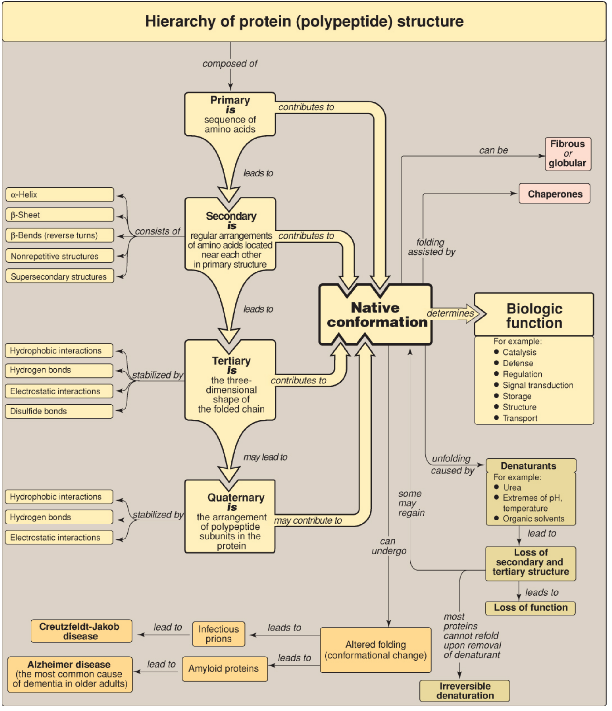
Figure 2.15 - Key concept map for protein structure. Quaternary structure is the arrangement of polypeptide subunits in the protein. (Lippincott Illustrated Reviews: Biochemistry, 8th Ed.)
Components: Subunits
Each polypeptide chain in a quaternary complex is called a subunit (also called a protomer). Subunit types are designated by Greek letters (α, β, γ, δ, etc.), with subscripts indicating the number of each type:
| Term | Meaning |
|---|---|
| Monomer | Single polypeptide chain - no quaternary structure |
| Homodimer | 2 identical polypeptide chains (α₂) |
| Heterodimer | 2 different polypeptide chains (αβ) |
| Homotetramer | 4 identical chains (α₄) |
| Heterotetramer | Mix of different chains (e.g., α₂β₂) |
For example, the notation α₂βγ denotes a protein with five subunits of three different types. - Harper's Illustrated Biochemistry, 32nd Ed.
Forces Stabilizing Quaternary Structure
Quaternary structure is held together primarily by noncovalent interactions at the interfaces between subunits. These are individually weak, but collectively very strong (like a Velcro fastener):
- Hydrophobic interactions - the dominant force; hydrophobic amino acid side chains are driven to the subunit interface, away from surrounding water
- Hydrogen bonds - between polar groups at the subunit contact surfaces
- Electrostatic (ionic/salt bridge) interactions - between oppositely charged residues (e.g., Asp/Glu vs. Lys/Arg/His)
- Disulfide bonds (S-S) - covalent bonds; some oligomeric proteins use interpolypeptide disulfide bonds between cysteine residues on different subunits to further strengthen the quaternary assembly
"Higher orders of protein structure are stabilized primarily - and often exclusively - by noncovalent interactions... collectively these individually weak but numerous interactions confer a high degree of stability upon the biologically functional conformation of a protein."
- Harper's Illustrated Biochemistry, 32nd Ed.
Classic Example: Hemoglobin
Hemoglobin is the textbook example of quaternary structure. It is a heterotetramer (α₂β₂) - composed of 2 alpha (α) and 2 beta (β) subunits. Other hemoglobin variants:
| Hemoglobin | Subunit Composition | Notes |
|---|---|---|
| HbA (normal adult) | α₂β₂ | Most common adult form |
| HbF (fetal) | α₂γ₂ | Higher O₂ affinity |
| HbS (sickle cell) | α₂β^S₂ | Point mutation in β chain |
| HbA₂ (minor adult) | α₂δ₂ | Minor fraction |
Why does quaternary structure matter for hemoglobin?
The quaternary structure of hemoglobin confers properties completely absent from the monomeric protein myoglobin:
- Cooperativity: binding of O₂ to one subunit increases the affinity of remaining subunits (sigmoidal O₂ binding curve vs. hyperbolic for myoglobin)
- Allosteric regulation: the T state (tense, low O₂ affinity) and R state (relaxed, high O₂ affinity) transitions arise from subunit rearrangements
- Bohr effect, 2,3-BPG binding, and CO₂ transport are all made possible by the tetrameric quaternary arrangement
"The quaternary structure of hemoglobin confers striking additional properties, absent from monomeric myoglobin, which adapts this tetrameric protein to its unique biologic roles in the reciprocal transport of O₂ and CO₂ between the lungs and peripheral tissues."
- Harper's Illustrated Biochemistry, 32nd Ed.
Visualization: All Four Levels
The diagram below shows all four levels of protein structure progressively, ending with quaternary structure (D - multiple subunits assembled in a membrane channel):
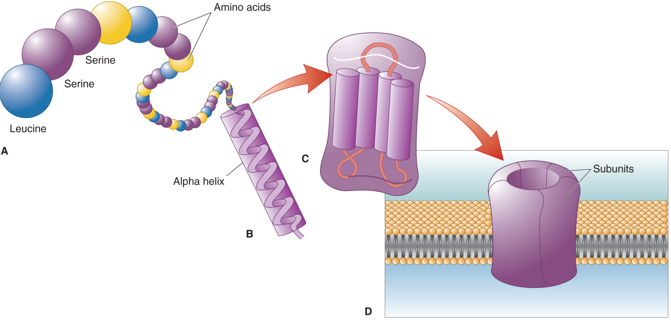
Figure 3.6 - Protein structure levels: (A) Primary, (B) Secondary (alpha helix), (C) Tertiary, (D) Quaternary - subunits assembled together. (Neuroscience: Exploring the Brain, 5th Ed.)
Other Key Examples of Quaternary Structure
| Protein | Subunit Composition | Function |
|---|---|---|
| Hemoglobin | α₂β₂ (tetramer) | O₂ transport |
| Collagen | Triple helix (3 chains) | Structural |
| Insulin | A + B chain dimer/hexamer | Hormone |
| Immunoglobulins (IgG) | 2 heavy + 2 light chains | Immunity |
| DNA polymerase | Multiple subunits | Replication |
| ATP synthase | ~8-10 subunits | Energy production |
Clinical Relevance
- Sickle cell disease: The single point mutation (Glu→Val) in the β-chain of HbS alters quaternary packing, causing HbS to polymerize when deoxygenated, deforming red blood cells.
- Thalassemias: Imbalanced synthesis of α or β chains disrupts normal quaternary assembly.
- Anti-GBM disease: Target epitopes lie in the quaternary structure of the α3 NC1 domain of collagen IV - the autoimmune attack is described as a "conformeropathy" involving perturbation of quaternary structure. - Harrison's Principles of Internal Medicine, 22nd Ed.
- Denaturants (urea, extremes of pH/temperature, organic solvents) can disrupt quaternary structure by breaking noncovalent interactions between subunits, leading to loss of function.
Sources: Lippincott Illustrated Reviews: Biochemistry 8th Ed. | Harper's Illustrated Biochemistry 32nd Ed. | Neuroscience: Exploring the Brain 5th Ed. | Harrison's Principles of Internal Medicine 22nd Ed.
in simple language for writing in exam
Quaternary Structure of Protein
(Exam-Ready Notes)
Definition
Quaternary structure is the arrangement of two or more polypeptide chains (called subunits) that join together to form one complete, functional protein.
Simply put: It is the "4th level" of protein structure where multiple chains come together.
Key Terms to Remember
- Subunit = each individual polypeptide chain in the complex
- Oligomeric protein = a protein with quaternary structure
- Protomer = the repeating structural unit
Forces That Hold Subunits Together
- Hydrophobic interactions - main stabilizing force
- Hydrogen bonds
- Ionic/Electrostatic interactions (salt bridges)
- Disulfide bonds - covalent; between cysteine residues of different chains (less common)
Note: Quaternary structure is stabilized primarily by non-covalent forces
Types of Quaternary Arrangement
| Type | Meaning | Example |
|---|---|---|
| Homodimer | 2 identical subunits (α₂) | - |
| Heterodimer | 2 different subunits (αβ) | - |
| Homotetramer | 4 identical subunits (α₄) | - |
| Heterotetramer | Mix of subunits (α₂β₂) | Hemoglobin (HbA) |
Best Example - Hemoglobin (Must Write in Exam!)
- Hemoglobin = α₂β₂ (2 alpha + 2 beta subunits) = heterotetramer
- Quaternary structure gives hemoglobin its cooperative O₂ binding (sigmoidal curve)
- Without quaternary structure (like myoglobin - only 1 chain), cooperative binding is lost
| Hemoglobin Type | Subunits | Notes |
|---|---|---|
| HbA (normal adult) | α₂β₂ | Most common |
| HbF (fetal) | α₂γ₂ | Higher O₂ affinity |
| HbS (sickle cell) | α₂β^S₂ | Mutant β chain |
Importance of Quaternary Structure
- Increases functional efficiency (e.g., cooperative binding in Hb)
- Allows allosteric regulation (T state ↔ R state in Hb)
- Provides structural stability
- Allows different subunits to carry out different functions in one complex
Clinical Points (for 1-2 marks)
- Sickle cell disease - mutation in β subunit → abnormal quaternary packing → HbS polymerizes → sickle shape
- Thalassemia - imbalanced subunit production → faulty quaternary assembly
- Denaturants (urea, extreme pH/heat) - break non-covalent bonds → quaternary structure disrupts → protein loses function
Quick One-liner for Exam
"Quaternary structure is the association of two or more polypeptide subunits held together by non-covalent interactions, as seen in hemoglobin (α₂β₂), which confers cooperative oxygen binding."
This is a shared conversation. Sign in to Orris to start your own chat.