
Granulomatous inflammation
granuloma histology multinucleated giant cells caseating
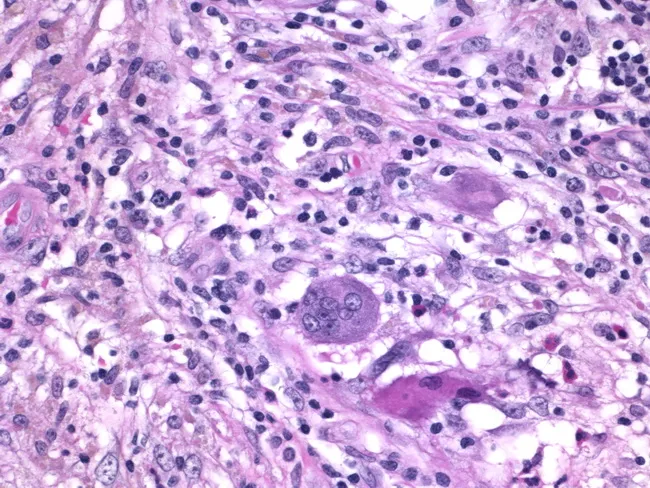
Histology of granulomatous mastitis on bright-field light microscopy. A dense inflammatory infiltrate forms granulomas centered on Langhans-type giant cells surrounded by epithelioid histiocytes, scattered lymphocytes, and occasional eosinophils. The image depicts a high-power view of the center of a granuloma within breast tissue biopsy; multinucleated giant cells with horseshoe-shaped nuclei are evident. The granulomatous architecture includes a peripheral rim of lymphocytes and histiocytes with minimal necrosis described. Staining by Hematoxylin and Eosin highlights basophilic nuclei and eosinophilic cytoplasm; pink collagenous stroma and adipose breast tissue may be visible in the background. Clinically, granulomatous mastitis can mimic infectious etiologies or neoplasia; the presence of Langhans giant cells strongly suggests a granulomatous process and warrants infectious workup for tuberculosis and fungal pathogens, as well as consideration of sarcoidosis or granulomatous reaction to foreign material. This image is diagnostic for granulomatous inflammation in breast tissue when correlated with clinical data and microbiologic studies; useful for education, differential diagnosis, and research on breast granulomatous disease. Additional educational value lies in distinguishing nonnecrotizing granulomas from caseating TB patterns, correlating histology with mammography and ultrasound, and guiding microbiologic testing and patient management in suspected granulomatous breast disease. The image supports teaching of granuloma biology and pathology.
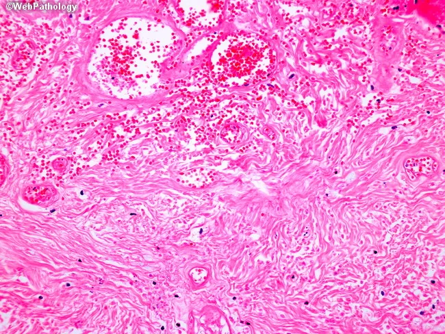
Light microscopy of an H&E-stained formalin-fixed tissue section at low to intermediate magnification reveals granulomatous inflammation characterized by well-formed nodular aggregates of epithelioid histiocytes and occasional Langhans-type multinucleated giant cells embedded in a fibrous stromal matrix. The granulomas appear in close association with accentuated lymphocytic cuffs and scattered plasma cells. Central areas show coagulative or caseating necrosis in some nodules, giving a pale eosinophilic center surrounded by a rim of epithelioid cells. The surrounding dermis/soft tissue shows delicate reticular fibers and vascular congestion with perivascular mononuclear infiltrates. There are no overt bacterial organisms evident with standard H&E, though organisms such as mycobacteria or fungi may require special stains (Ziehl-Neelsen, GMS) to confirm infection. The architecture suggests a granulomatous process rather than acute suppurative inflammation. Diagnostic significance: indicative of granulomatous disease; differential includes infectious etiologies (tuberculosis, fungal infections) and noninfectious conditions (sarcoidosis, foreign-body reaction). Clinical correlation recommended with exposure history, immunologic status, and targeted staining/cultures. Potential use cases: educational demonstration of granuloma morphology, pathology student review, differential diagnosis in infectious and granulomatous diseases, histology atlas reference. This description supports search for terms granuloma, epithelioid histiocytes, Langhans giant cells, caseation, TB, sarcoidosis, histopathology.
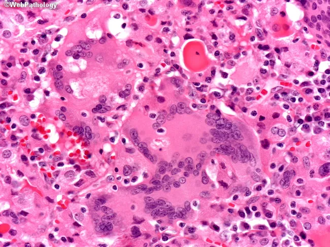
High-magnification bright-field histology of thyroid gland tissue stained with Hematoxylin and Eosin (H&E). The specimen shows granulomatous inflammation characterized by numerous foreign body giant cells and aggregates of epithelioid histiocytes embedded in a pink cytoplasm background. The thyroid parenchyma is variably disrupted with scattered lymphocytes and plasma cells; there is no well-formed granuloma rim, and there is no focal caseous necrosis evident. Multinucleated giant cells frequently contain cholesterol-rich debris and refractile material consistent with a foreign body reaction. The microarchitecture lacks overt cytologic atypia and no malignant cells are seen. Follicular colloid appears diminished in areas of inflammation. These features are compatible with granulomatous thyroiditis, most consistent with subacute (de Quervain-like) thyroiditis, but the absence of caseation argues against tuberculous thyroiditis. The image highlights the distinction between inflammatory thyroid disease and infectious etiologies. Clinically, such histology correlates with tender thyroid, transient thyrotoxic phase followed by hypothyroidism in some cases, elevated ESR, and leukocytic infiltration. Differential diagnoses include sarcoid thyroiditis and TB, which would show caseating granulomas or granulomatous inflammation with caseation. This high-magnification view is valuable for educational purposes and for correlating histology with endocrine pathology. Clinicians should correlate clinically with serology and imaging. Useful for teaching and research.
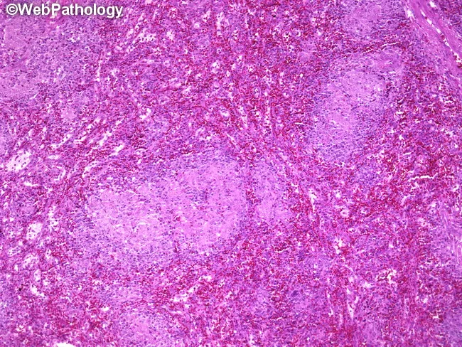
This is a photomicrograph of a splenic parenchyma section from a splenectomy specimen, prepared for light microscopy and stained with hematoxylin and eosin. The specimen shows multiple subcentimeter nodules within the splenic parenchyma, with well-formed, non-caseating granulomas composed of tightly arranged epithelioid macrophages and multinucleated giant cells surrounded by a lymphocytic rim. The granulomas lack central necrosis, which, along with histiocytic microarchitecture, is highly suggestive of granulomatous inflammation due to Toxoplasma gondii infection. Serologic tests confirmed toxoplasmosis in this patient, correlating with disseminated disease or focal splenic involvement associated with lymphadenopathy and splenomegaly. In the spleen, toxoplasmosis can produce discrete nodules or focal granulomatous lesions that may mimic lymphoma or other granulomatous diseases on imaging or gross examination. Key differential diagnoses include sarcoidosis, fungal infections such as histoplasmosis, mycobacterial infections, and other parasitic infestations; clinical correlation with serology and immune status is essential. This image is relevant for teaching granulomatous splenitis due to toxoplasmosis, assessing granuloma morphology, distinguishing non-caseating from caseating granulomas, and correlating histology with systemic infection. Appropriate clinical scenarios include immunocompromised patients with fever, cytopenias, or organomegaly and routine pathology review in splenectomy specimens. This image exemplifies infectious granulomatous disease companion to serology and morphology in clinical practice.
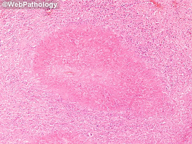
This is a low-magnification brightfield histology image of a tissue section stained with Hematoxylin and Eosin (H&E). The specimen shows granulomatous inflammation with aggregates of epithelioid macrophages forming a rounded granuloma, often with multinucleated giant cells, surrounded by a lymphocytic cuff. The central focus is well circumscribed within native parenchyma, suggesting a chronic, organized immune response to a persistent antigen. Necrosis is not clearly evident at this magnification, though subtle caseation cannot be excluded. Morphology is compatible with tuberculoid or non-caseating granulomas, and infectious versus noninfectious etiologies must be distinguished with ancillary studies. This pattern can occur in lymph nodes or solid organs and prompts a differential that includes tuberculous lymphadenitis, sarcoidosis, fungal granulomatous infections (Histoplasma, Coccidioides), and foreign-body reaction. Clinically, recognition of granulomas guides testing: acid-fast bacilli staining (Ziehl-Neelsen), fungal stains (GMS/PAS), cultures, PCR panels, and radiologic correlation. The image is educational for medical trainees, illustrating granuloma architecture, macrophage differentiation, and the spectrum of chronic inflammatory responses. Educational context.
granulomatous inflammation pathogenesis
Granulomatous Inflammation
- Epithelioid cells: activated macrophages develop abundant pink granular cytoplasm with indistinct borders, resembling epithelial cells (hence "epithelioid").
- Multinucleated giant cells: formed by fusion of multiple activated macrophages, typically 40-50 μm in diameter. When the nuclei are arranged peripherally in a horseshoe pattern they are called Langhans giant cells; giant cells engulfing foreign material with haphazardly scattered nuclei are called foreign-body giant cells.
- Epithelioid cell aggregates are usually rimmed by a collar of lymphocytes; older granulomas develop a peripheral rim of fibroblasts and connective tissue.
- In infections such as Mycobacterium tuberculosis, hypoxia and free-radical-mediated injury produce a central zone of caseous necrosis - grossly granular and cheese-like, microscopically amorphous and eosinophilic with loss of cellular detail - Robbins & Kumar Basic Pathology, p. 106.
- Immune (delayed-type hypersensitivity) granulomas: caused by persistent, poorly degradable antigens (usually microbes) that drive a sustained T-cell-mediated immune response. Th1 cells secrete IFN-gamma, which is the principal macrophage-activating cytokine and central to granuloma formation. In some parasitic infections (e.g., schistosomiasis) a Th2-driven response predominates, with eosinophil-rich granulomas.
- This image below shows non-caseating granulomas from disseminated toxoplasmosis, illustrating an immune granuloma without necrosis:
- Foreign-body granulomas: reactions to inert, non-immunogenic material that is simply too large for a single macrophage to phagocytose (e.g., talc from intravenous drug use, suture material, other fibers). No T-cell-mediated immune response is required. Epithelioid cells and giant cells appose directly to the surface of the material, which is often visible in the center of the granuloma - and appears refractile under polarized light.
| Category | Examples |
|---|---|
| Mycobacterial | Tuberculosis (caseating), leprosy, atypical mycobacteria |
| Fungal | Histoplasmosis, coccidioidomycosis, cryptococcosis (all can caseate) |
| Bacterial/spirochetal | Cat-scratch disease (Bartonella), syphilis (gumma), tularemia |
| Parasitic | Schistosomiasis |
| Idiopathic/immune-mediated | Sarcoidosis (non-caseating, "naked" granulomas), Crohn disease, granulomatosis with polyangiitis |
| Foreign body | Talc, suture material, silica, beryllium |
| Other organ-specific | Granulomatous mastitis, subacute (de Quervain) thyroiditis |