
Shock
"shock" AND "management"
shock types classification pathophysiology diagram
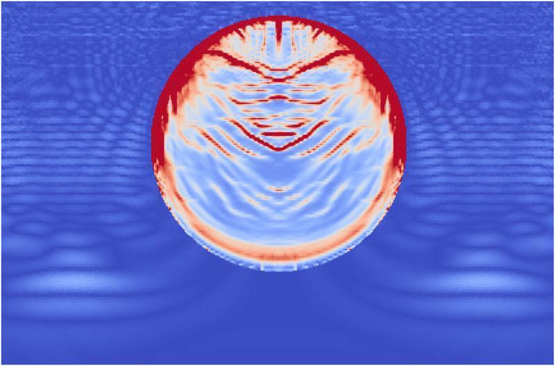
This pathophysiology diagram is a computational visualization using the Material Point Method (MPM) to simulate the effects of Extracorporeal Shock Wave Lithotripsy (ESWL) on a kidney stone. The image depicts a circular, single-phase kidney stone immersed in a fluid medium (modeled as water) at the moment a high-pressure shock wave has reached the end of the stone. The fluid medium displays stress waves as concentric, blue-toned undulating ripples propagating through the domain. Within the stone, damage accumulation is color-coded: red indicates areas of complete failure, while lighter blue and white areas represent internal stress and emerging fractures. Significant damage is concentrated at the proximal side (top) where the wavefront first impacted, showing a thick layer of failure. Distinct angled failure lines extend from the stone's edges toward the center, representing shear stress interactions. Minor damage and tensile fractures are also visible at the distal end (bottom) of the stone, resulting from internal wave reflections and interference patterns. This simulation serves as an educational model for understanding fragmentation mechanisms in urolithiasis treatment.
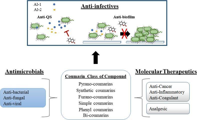
This medical pathophysiology diagram illustrates the multi-functional pharmacological properties of the coumarin class of compounds, focusing on their role as anti-infectives. The schematic is divided into two primary sections: a broad classification of coumarin types and their therapeutic applications, and a detailed mechanistic view of their anti-infective properties. The lower half categorizes coumarins into various structural types (pyrano-coumarins, synthetic, furano-coumarins, etc.) and links them to 'Antimicrobials' (anti-bacterial, anti-fungal, anti-viral) and 'Molecular Therapeutics' (anti-cancer, anti-inflammatory, anti-coagulant, analgesic). The upper section details the 'Anti-infective' mechanism, specifically highlighting 'Anti-QS' (quorum sensing) and 'Anti-biofilm' activities. In the 'Anti-QS' portion, coumarin structures are shown inhibiting signaling molecules AI-1 (blue) and AI-2 (yellow), represented by T-bar inhibition icons between bacteria. In the 'Anti-biofilm' portion, a red cross over a transition arrow signifies the inhibition of individual bacterial cells aggregating into a multicellular biofilm structure. This educational infographic summarizes how coumarins act as non-toxic modulators of microbial behavior by disrupting cell-to-cell communication and structural development in pathogens.
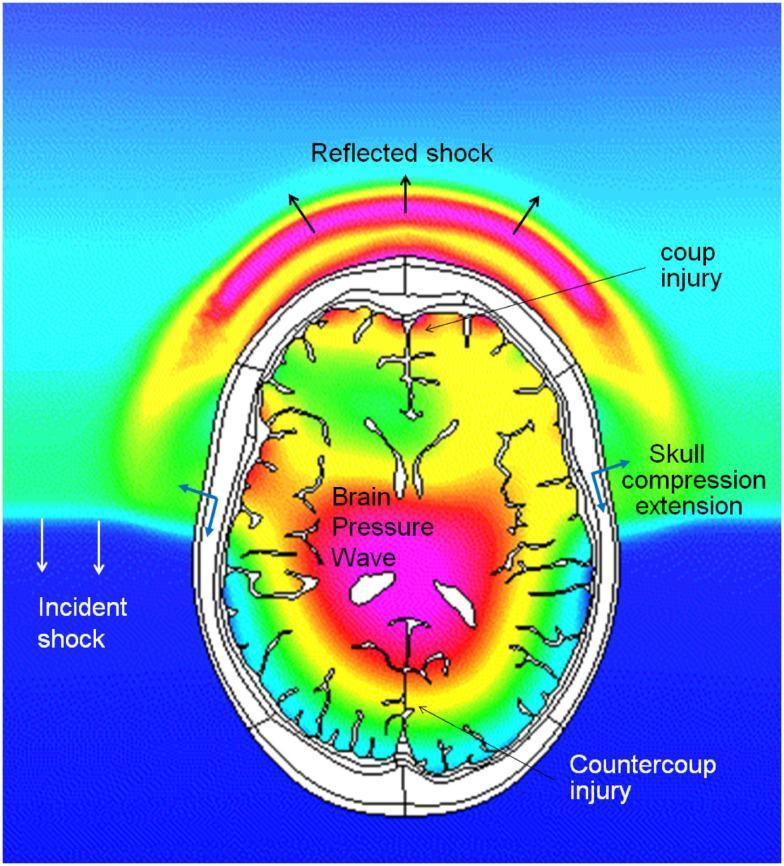
This pathophysiology diagram depicts a coupled simulation of Computational Fluid Dynamics (CFD) and Finite Element Method (FEM) biomechanics, illustrating a blast wave's interaction with a human head. The visualization shows an 'Incident shock' wave impacting the inferior-lateral aspect of the skull, resulting in a 'Reflected shock' wave arcing away from the impact site. The diagram highlights critical injury mechanisms including 'coup injury' at the site of impact and 'countercoup injury' on the diametrically opposite side of the brain. A color-coded pressure gradient within the cranial cavity shows the 'Brain Pressure Wave' propagating through the tissue, with high-pressure regions indicated in red/pink and lower-pressure zones in green. Structural mechanical responses are noted by labels for 'Skull compression extension.' This content is designed for advanced medical education in neurology and trauma surgery to explain the complex biomechanics of blast-induced traumatic brain injury (bTBI) and intracranial pressure dynamics.
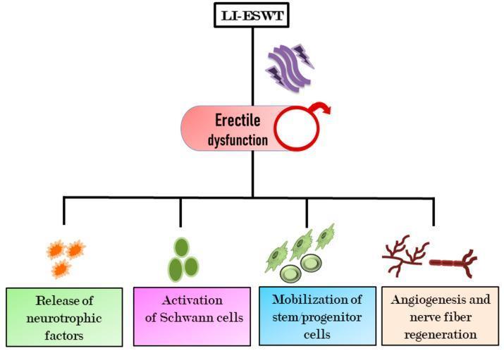
This pathophysiology diagram illustrates the therapeutic mechanisms of Low-Intensity Extracorporeal Shock Wave Therapy (LI-ESWT) in the treatment of erectile dysfunction (ED). The central flowchart displays LI-ESWT as the primary intervention, targeting a node labeled 'Erectile dysfunction' marked with a male symbol. Downstream of this interaction, the diagram identifies four key regenerative processes stimulated by shockwave energy: 1) Release of neurotrophic factors (depicted by orange star-burst icons), 2) Activation of Schwann cells (represented by green ovoid cell icons), 3) Mobilization of stem/progenitor cells (illustrated as green undifferentiated cells), and 4) Angiogenesis and nerve fiber regeneration (visualized as red branching vascular and neural structures). The diagram summarizes the biological pathways by which LI-ESWT promotes tissue repair, neovascularization, and improved hemodynamics within the penile corpora cavernosa. It is designed as an educational overview of regenerative medicine and urological therapy for medical students and clinicians.
Shock
Definition
Classification
| Type | Key Examples | Core Mechanism |
|---|---|---|
| Cardiogenic | MI, ventricular rupture, arrhythmia, cardiac tamponade, pulmonary embolism | Pump failure - reduced cardiac output from intrinsic or extrinsic causes |
| Hypovolemic | Hemorrhage, severe burns, vomiting, diarrhea | Inadequate blood or plasma volume leading to low preload and low CO |
| Septic | Overwhelming bacterial/fungal infection | Cytokine cascades; peripheral vasodilation; endothelial injury; DIC |
| Neurogenic | Spinal cord injury, high spinal anesthesia | Loss of sympathetic tone → acute vasodilation → hypotension |
| Anaphylactic | IgE-mediated hypersensitivity | Acute vasodilation from mast cell degranulation |
| Obstructive | Pulmonary embolism, aortic dissection, pericardial tamponade | Mechanical obstruction to cardiac output |
- Robbins, Cotran & Kumar, p. 134 (Table 4.3)
- Goodman & Gilman's Pharmacological Basis of Therapeutics, p. 285
Hemodynamic Profiles
| Parameter | Cardiogenic | Hypovolemic | Septic (early) | Neurogenic |
|---|---|---|---|---|
| CO/CI | ↓↓ | ↓ | ↑ (high output) | ↓ |
| SVR | ↑↑ | ↑ | ↓↓ | ↓↓ |
| CVP/PCWP | ↑ | ↓↓ | ↓ | ↓ |
| Skin | Cold/clammy | Cold/clammy | Warm/flushed (early) | Warm |
| Pulse | Thready/weak | Thready/weak | Bounding (early) | Bradycardia possible |
Pathophysiology: Stages of Shock
1. Compensated (Nonprogressive) Stage
- Baroreceptor reflexes → sympathetic activation
- Catecholamine release → tachycardia, vasoconstriction
- ADH release → renal water retention
- RAAS activation → aldosterone, Na+ retention
- Net effect: blood is shunted from skin/kidneys to heart and brain; cool, pale skin; oliguria. In septic shock early on, peripheral vasodilation may cause warm, flushed skin instead.
2. Progressive Stage
- Aerobic respiration → anaerobic glycolysis → lactic acidosis
- Lactic acidosis blunts the vasomotor response → arteriolar dilation → blood pools in the microcirculation
- Peripheral pooling further worsens CO and predisposes endothelium to anoxic injury and DIC
- Vital organs (heart, brain, kidneys) begin to fail
3. Irreversible Stage
- Lysosomal enzyme leakage worsens cellular injury
- Myocardial contractility declines (partly from increased NO synthesis)
- Ischemic bowel allows intestinal flora into circulation → superimposed bacteremia
- Acute tubular necrosis → renal failure
- Despite intervention, death follows
- Robbins, Cotran & Kumar, p. 136
Septic Shock: Pathogenesis in Detail
-
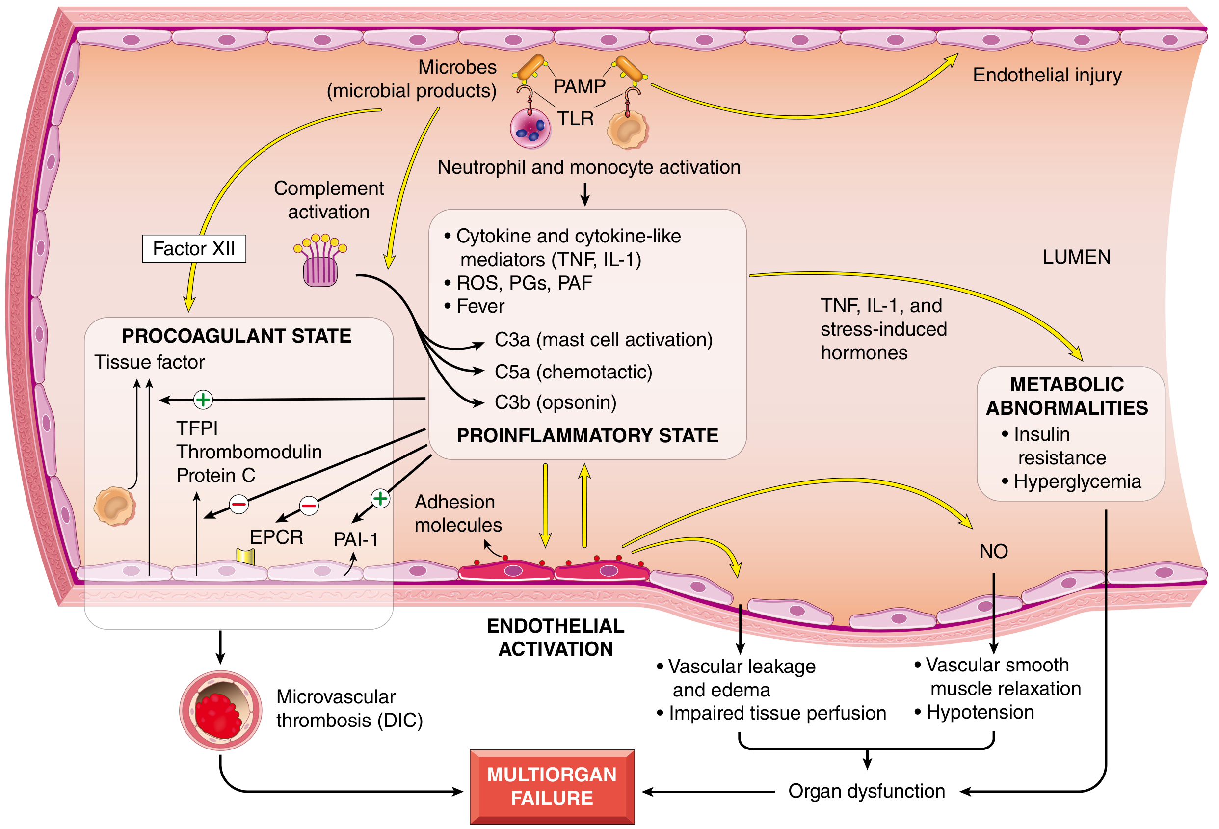
Proinflammatory cascade: Microbial PAMPs/DAMPs → TLR activation → NF-κB → TNF, IL-1, IL-12, IL-18, IFN-γ, HMGB1, ROS, prostaglandins, PAF → complement activation (C3a, C5a, C3b)
-
Counter-inflammatory immunosuppression: Shift from Th1 to Th2 cytokines; IL-10 production; lymphocyte apoptosis → patients oscillate between hyperinflammatory and immunosuppressed states
-
Endothelial activation/injury: Inflammatory cytokines loosen tight junctions → vascular leakage, edema → impaired perfusion; upregulation of NO → vasodilation and systemic hypotension; loss of capillary autoregulation
-
Procoagulant state: ↑ Tissue factor on monocytes + ↓ anticoagulant factors (thrombomodulin, protein C, TFPI) + ↑ PAI-1 → systemic thrombin activation → fibrin-rich microthrombi → DIC in up to 50% of septic patients
-
Metabolic abnormalities: Insulin resistance, hyperglycemia (via TNF/IL-1, glucocorticoids, catecholamines), ↑ triglycerides and lactate; possible adrenal insufficiency (Waterhouse-Friderichsen syndrome in DIC)
Morphology (Organ-Level Effects)
- Kidney: Fibrin thrombi in glomeruli; acute tubular necrosis
- Adrenals: Cortical lipid depletion (increased steroid synthesis) or frank necrosis (DIC)
- Lungs: Resistant in pure hypovolemic shock, but sepsis/trauma → diffuse alveolar damage ("shock lung" / ARDS)
- Brain, heart, GI tract: Also commonly affected
- Recovery is possible except for neuronal and cardiomyocyte loss
Clinical Features
| Feature | Hypovolemic/Cardiogenic | Septic (early) |
|---|---|---|
| BP | Hypotension | Hypotension |
| Pulse | Weak, rapid | Bounding (later weak) |
| Skin | Cool, clammy, cyanotic | Warm, flushed |
| Breathing | Tachypnea | Tachypnea |
| Urine output | Oliguria | Variable |
| Mental status | Confusion, obtundation | Confusion |
Management
General Principles
- Airway and breathing - ensure oxygenation
- Identify and treat the underlying cause simultaneously
- Maintain/restore blood volume with hemodynamic monitoring
- Vasoactive drugs if volume alone is insufficient
Fluid Resuscitation
- Isotonic crystalloid (Normal Saline or Lactated Ringer's) - note only ~175-200 mL of a 1 L infusion remains intravascular after a few hours
- For hemorrhagic shock: blood transfusion preferred over crystalloid beyond 1-2 L, as it replaces both volume and oxygen-carrying capacity
- Avoid routine pulmonary artery catheters (no longer recommended)
Vasoactive/Inotropic Drugs
- Dopamine: Effective inotrope with less tachycardia than isoproterenol; low doses promote renal dilation; high doses (>10-20 mcg/kg/min) → α-receptor activation → vasoconstriction
- Dobutamine: Preferred in cardiogenic shock - increases contractility with minimal tachycardia or SVR change; best for optimizing preload/afterload/contractility
- Norepinephrine: Strong α-agonist - used when vasopressor support is needed; risk of renal/gut ischemia in cardiogenic shock
- Epinephrine: Increases HR and contractility but may provoke arrhythmias
- Vasodilators (nitroprusside): In cardiogenic shock where reflex vasoconstriction is already intense, reducing afterload may improve CO more than vasopressors
Specific Therapy by Type
- Hypovolemic: Stop bleeding (surgical if needed), volume replacement with blood products
- Cardiogenic: Reperfusion (PCI for STEMI), inotropes, IABP or mechanical circulatory support
- Septic: Source control (drain abscess, remove lines), early antibiotics (prolonged/continuous β-lactam infusions shown to improve outcomes in meta-analysis [PMID 38864162]), vasopressors (NE first-line)
- Obstructive: Remove obstruction (fibrinolysis/embolectomy for PE; pericardiocentesis for tamponade)
- Anaphylactic: Epinephrine IM (first-line), fluids, antihistamines, steroids
Prognosis
- Hypovolemic shock in otherwise healthy young patients: >90% survival with appropriate management
- Septic or cardiogenic shock: Substantially worse outcomes even with optimal care
- Robbins, Cotran & Kumar, p. 137
Recent Evidence (2024-2026)
- A 2024 meta-analysis in JAMA (PMID 38864162) found that prolonged/continuous β-lactam infusions (vs. intermittent) in sepsis/septic shock reduce mortality - a practice shift worth noting.
- The 2026 Surviving Sepsis Campaign pediatric guidelines (PMID 41869844) provide updated management recommendations for children with septic shock.