
Cervical disc replacement ms ortho 20 marks answer
Reading File
Now let me search the library for cervical disc replacement content in parallel with a web search.
Finding Sources
Finding Sources
Searching PubMed
"cervical disc arthroplasty" OR "cervical disc replacement"
I found the content in Campbell's. Let me read the full cervical disc arthroplasty section.
Reading File
Excellent content. Let me read more of this section for completeness.
Reading File
I went past the CDA section. Let me go back to the correct offset range to read the CDA section content.
Reading File
Reading File
Reading File
I now have comprehensive content from Campbell's Operative Orthopaedics 2026. Let me also fetch a couple of the device images to include in the answer.
Loading Image
Loading Image
I now have all the material needed for a thorough MS Ortho 20-mark answer. Let me compose it.
Cervical Disc Replacement (Cervical Disc Arthroplasty)
MS Orthopaedics — 20 Marks
1. Introduction and Rationale
Cervical disc replacement (CDR), also called cervical disc arthroplasty (CDA), is a motion-preserving alternative to anterior cervical discectomy and fusion (ACDF) for symptomatic cervical disc disease. The fundamental rationale is that by avoiding solid fusion, the abnormal stresses transferred to adjacent motion segments (adjacent segment disease/ASD) can be minimized, thereby reducing the need for reoperation at adjacent levels. Multiple randomized controlled trials have confirmed that CDA achieves at least equivalent neurological outcomes to ACDF while maintaining segmental motion and producing faster return to work (approximately 2 weeks earlier).
(Campbell's Operative Orthopaedics, 15th Ed 2026, p. 2310)
2. Anatomy Relevant to the Procedure
The anterior cervical spine is accessed via the standard Smith-Robinson approach. Key anatomical relationships:
- Anterior cervical triangle - bordered by sternocleidomastoid laterally, trachea and esophagus medially
- Carotid sheath - retracted laterally; contains carotid artery, internal jugular vein, vagus nerve
- Recurrent laryngeal nerve - at risk with retraction; runs in the tracheoesophageal groove (left side more reliable, preferred side for approach by some surgeons)
- Sympathetic chain - lies on the longus colli; vigorous lateral retraction causes Horner's syndrome
- Disc space - C5-6 and C6-7 are most commonly affected. The uncinate processes define the lateral extent of disc removal; they must be preserved for foraminal decompression and as a guide for implant sizing
- Vertebral artery - lies in the transverse foramen of C6 and above; at risk if uncinate process is breached
3. Indications (Box 45.8, Campbell's)
- Symptomatic cervical disc disease at one or two levels between C3 and T1
- Confirmed on MRI/CT/myelography showing herniated nucleus pulposus, spondylosis, or loss of disc height
- Failed ≥6 weeks of conservative therapy
- Age 20-70 years
- No contraindications present
The indications are essentially the same as for ACDF - radiculopathy from disc herniation or spondylosis, and myelopathy from single or two-level disease.
4. Contraindications
Absolute:
| Category | Details |
|---|---|
| Level extent | ≥3 vertebral levels requiring treatment |
| Instability | Sagittal translation >3 mm and/or >11-degree rotational difference vs. adjacent level |
| Allergy | Titanium, polyethylene, cobalt, chromium, or molybdenum |
| Adjacent fusion | Pre-existing fusion adjacent to the planned level |
| Bone quality | Osteoporosis / osteopenia; other metabolic bone disease (Paget's, osteomalacia) |
| Facet disease | Significant facet joint degeneration at the target level |
| Severe spondylosis | Bridging osteophytes, disc height loss >50%, motion <2 degrees |
| Prior surgery | Previous surgery at the same level |
| Deformity | Post-traumatic vertebral body deficiency |
| Axial pain only | Neck pain as the sole symptom with no neural compromise |
| Infection | Active local or systemic infection |
| Malignancy | Active or within 5 years |
| Systemic disease | HIV, AIDS, hepatitis B/C, insulin-dependent diabetes |
| Obesity | BMI >40 kg/m² or >100 lbs over ideal body weight |
| Pregnancy | Pregnant or planning pregnancy in next 3 years |
| Medications | Corticosteroids or drugs interfering with bone/soft tissue healing |
| Autoimmune | Rheumatoid arthritis, ankylosing spondylitis |
Additional anatomic consideration: Facet arthroplasty treats only one of the three joints at each motion segment. CDA is therefore inappropriate when CT shows significant facet overload and degeneration. OPLL (ossification of posterior longitudinal ligament) and coexistent posterior compressive pathology are also contraindications.
5. Pre-operative Workup
- X-rays (AP, lateral, flexion-extension): Assess instability, disc height, lordosis, existing osteophytes, motion at the target level (must show ≥2 degrees to qualify)
- MRI: Confirms level, quantifies cord/root compression, assesses disc hydration, evaluates facet joints
- CT scan: Evaluates extent of spondylosis, bony anatomy, foraminal stenosis, OPLL
- DEXA scan: Rules out osteoporosis (T-score ≥ -1.0 required)
- Neurological assessment: NDI (Neck Disability Index), VAS pain score, mJOA for myelopathy
6. Devices Available
Multiple devices have received FDA approval. They differ in articulation design and materials:
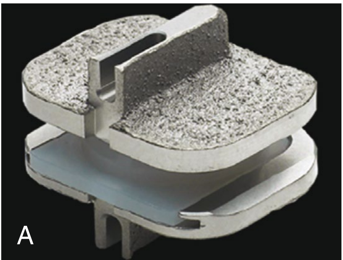
Fig. A - Prestige LP / PCM type device (metal endplates with polyethylene core)
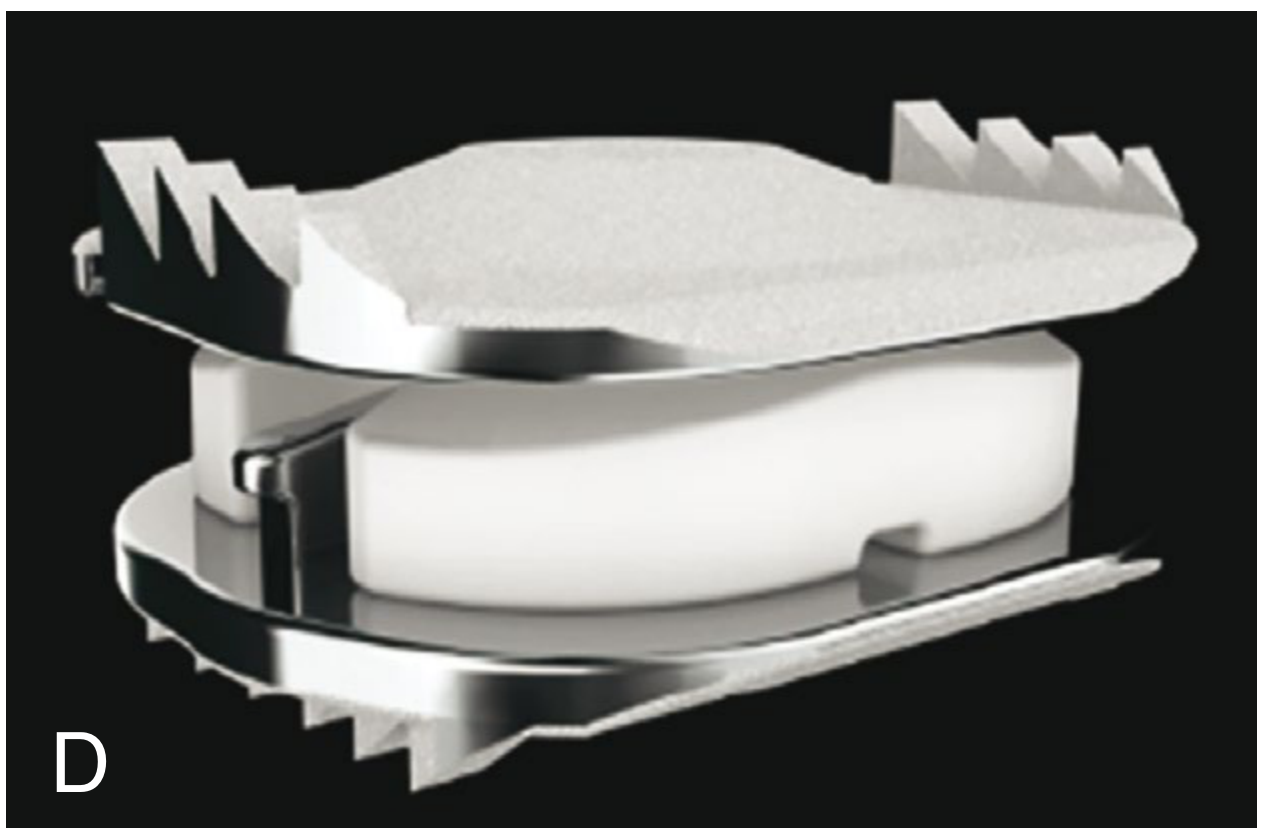
Fig. B - Mobi-C type device (semi-constrained mobile core)
FDA-Approved Devices (Table 45.4, Campbell's 2026):
| Device | Manufacturer |
|---|---|
| Prestige Cervical Disc System | Medtronic Sofamor Danek |
| Bryan Cervical Disc | Medtronic Sofamor Danek |
| ProDisc-C | Synthes Spine |
| Mobi-C Cervical Disc Prosthesis | ZimVie |
| PCM Cervical Disc | NuVasive |
| Secure-C Artificial Cervical Disc | Globus Medical |
| ActivL Artificial Disc | B. Braun Aesculap |
Design types:
- Constrained (ball-in-socket): ProDisc-C, Prestige - fixed center of rotation
- Semi-constrained / mobile core: Bryan, Mobi-C - allow translation + rotation
- Unconstrained: Allow 6 degrees of freedom; approximate normal disc kinematics
Materials:
- Metal-on-metal (cobalt-chrome alloy): Prestige
- Metal-on-polyethylene: ProDisc-C, Mobi-C, Secure-C
- Polymer core (polyurethane + titanium endplates): Bryan
7. Surgical Technique
Approach
- Standard anterior Smith-Robinson approach, usually right-sided
- Patient supine, neck slightly extended (Gardner-Wells traction or head halter optional)
- Longitudinal incision along the medial border of sternocleidomastoid
Steps
- Exposure: Blunt dissection through the platysma; carotid sheath retracted laterally, trachea/esophagus medially; longus colli elevated laterally to expose the disc
- Level confirmation: C-arm fluoroscopy with spinal needle
- Disc removal: Complete anterior discectomy; ALL sectioned carefully (unlike ACDF, some motion must be preserved)
- End-plate preparation: Distraction maintained with Caspar pins; cartilaginous endplates removed down to cortical bone using curettes and rasps - cortical bone must be preserved (unlike fusion, no bone grafting)
- Decompression: Posterior osteophytes removed with high-speed burr/Kerrison rongeurs; bilateral foraminal decompression performed
- Sizing and trialing: Template used; prosthesis must span as much endplate area as possible to distribute load evenly
- Implantation: Device inserted and impacted/screwed into place; position confirmed with fluoroscopy in both planes
- Ideal position (Fig. 45.18):
- Height similar to adjacent normal discs
- Maximum endplate coverage
- Centrally positioned in both sagittal and coronal planes
Intraoperative pitfalls
- Undersized implant → stress concentration per unit area → subsidence; inadequate endplate coverage → heterotopic ossification and osteophytes → loss of motion
- Malpositioning (coronal plane) → unilateral foraminal narrowing → radiculopathy
- Overstuffing (oversized implant) → facet joint distraction → axial neck pain, referred scapular pain, reduced segmental motion
8. Advantages Over ACDF
| Parameter | CDA | ACDF |
|---|---|---|
| Motion preservation | Yes | No (fused) |
| Adjacent segment disease | Reduced | 2.9% per year (cumulative) |
| Re-operation rate | Lower at 10 years | Higher |
| Return to work | ~2 weeks earlier | Standard |
| Axial neck pain | Less | More (donor site + fusion mass) |
| Collar / bracing | Not required | Often used |
| Number of levels | 1-2 levels (best) | 1 to multi-level |
9. Results and Evidence
Long-term randomized controlled trial data (7-10 years follow-up) consistently show CDA to be superior or non-inferior to ACDF:
- NDI scores and VAS neck/arm pain equivalent or better with CDA
- Reoperation at the index level lower with CDA
- Secondary surgery at adjacent levels significantly lower with CDA
- Motion preservation better maintained with arthroplasty vs arthrodesis
- NORCAT trial (Norwegian Cervical Arthroplasty Trial): CDA = ACDF at 2 years for single-level disease
Recent meta-analyses (2025) confirm CDA superiority over ACDF for single-level disease in NDI, neurological success rate, and adjacent segment reoperation at minimum 4-year follow-up (PMID 40797275), and for cervical spondylotic myelopathy (PMID 40184678).
10. Complications
| Complication | Notes |
|---|---|
| Implant migration / subsidence | Undersized prosthesis; poor bone quality |
| Heterotopic ossification (HO) | Most common complication; causes motion loss; graded by McAfee classification |
| Recurrent radiculopathy | From HO, retained osteophyte, or foraminal narrowing from malposition |
| Adjacent segment degeneration | Less than ACDF but not eliminated |
| Dysphagia | Retractor-related; usually temporary |
| Hoarseness / RLN palsy | Recurrent laryngeal nerve traction |
| Horner's syndrome | Sympathetic chain injury |
| Vertebral artery injury | Rare; catastrophic; from lateral breach; systematic review PMID 40544643 |
| Metal-on-metal reaction | Lymphocytic reaction similar to failed MoM hip prostheses; rare but described |
| Device failure / breakage | Rare with modern implants |
| C5 palsy | Root traction during decompression |
| Wound infection / hematoma | General surgical risk |
Heterotopic Ossification (HO) deserves special emphasis:
- The most common late complication
- Graded 0-4 (McAfee classification):
- Grade 0: No HO
- Grade 1: HO outside disc space
- Grade 2: HO within disc space without bridging
- Grade 3: HO bridging with preserved motion
- Grade 4: Complete fusion (ankylosis)
- Grade 3-4 HO eliminates the motion-preserving benefit of CDA
- Risk factors: inadequate endplate coverage, undersized implant, retained posterior osteophytes
11. Post-operative Management
- Soft collar for comfort only (not mandatory)
- Early mobilization and physiotherapy
- No rigid immobilization (unlike ACDF)
- Activity restrictions: avoid contact sports for 6 weeks
- Follow-up radiographs at 6 weeks, 3 months, 6 months, 1 year - assess motion, HO formation, implant position
- NDI and VAS scoring at each visit
12. Special Considerations
- Two-level CDA (C4-5 and C5-6, or C5-6 and C6-7): FDA-approved for Mobi-C; evidence shows superiority over two-level ACDF in reoperation rates
- CDA vs posterior foraminotomy: CDA preferred when anterior pathology predominates; posterior foraminotomy for soft lateral disc herniation is an equally effective, less invasive option
- Facet condition: CT mandatory pre-operatively; significant facet degeneration is an absolute contraindication as CDA treats only the disc joint (one of three joints in the motion segment)
- Bone quality: DEXA scan mandatory; osteoporosis predisposes to endplate subsidence and HO
13. Summary
Cervical disc replacement is a well-established surgical procedure for one or two-level cervical disc disease causing radiculopathy or myelopathy that has failed conservative management. Its primary advantage over ACDF is preservation of motion and reduction of adjacent segment disease. Careful patient selection (ruling out facet degeneration, instability, osteoporosis, OPLL) and meticulous surgical technique - particularly achieving the correct implant size and central positioning - are the determinants of long-term success. The evidence from multiple 10-year RCTs and recent meta-analyses supports non-inferiority or superiority of CDA over ACDF across clinical, radiological, and reoperation outcomes.
Source: Campbell's Operative Orthopaedics, 15th Edition 2026, Chapter 45 - Cervical Disc Arthroplasty, pp. 2310-2312. Recent evidence: Zhang et al., J Orthop Surg Res 2025 (PMID 40797275); Huang et al., J Neurosurg Spine 2025 (PMID 40184678).
This is a shared conversation. Sign in to Orris to start your own chat.