
antihistamines
antihistamine drug classification H1 H2 receptor diagram
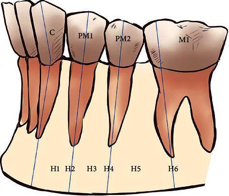
This anatomical diagram provides a schematic representation of the horizontal classification of the mental foramen (MF) position relative to the mandibular teeth, commonly used in maxillofacial radiology and surgical planning. The illustration depicts the left mandibular segment from the canine to the first molar. Six specific horizontal positions (H1–H6) are categorized based on their anterior-posterior relationship to the tooth axes: H1 is located between the canine (C) and first premolar (PM1); H2 aligns with the long axis of the PM1; H3 is positioned between the first and second premolars (PM2); H4 aligns with the long axis of the PM2; H5 is located between the PM2 and the first molar (M1); and H6 aligns with the mesial root of the M1. Blue vertical lines illustrate the long axes of the teeth to define these anatomical landmarks. This diagram serves as an educational tool for dental students and clinicians to understand the common anatomical variations of the mental nerve exit point, which is critical for local anesthesia and avoiding neurovascular injury during dental implantation or mandibular surgery.
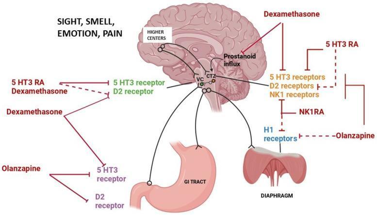
A pathophysiology diagram and pharmacological algorithm illustrating the mechanisms of chemotherapy-induced nausea and vomiting (CINV) and the corresponding sites of action for anti-emetic agents. The central anatomical figure is a sagittal section of the human brain, highlighting the Higher Centers (processing sensory/emotional stimuli), the Vomiting Center (VC) in the medulla, and the Chemoreceptor Trigger Zone (CTZ). Neural pathways connect these central regions to peripheral effectors including the GI tract and the diaphragm. The diagram maps specific drug classes to their molecular targets: 5-HT3 Receptor Antagonists (5-HT3 RA), NK1 Receptor Antagonists (NK1RA), Dexamethasone, and Olanzapine. These agents are shown inhibiting various receptors including 5-HT3, D2 (dopamine), NK1 (neurokinin-1), and H1 (histamine) receptors within the CTZ and peripheral pathways. Dexamethasone is specifically noted for its role in inhibiting prostanoid influx. Solid red lines represent predominant mechanisms of action, while dashed lines indicate minor pathways. This educational visual summarizes multi-receptor antagonism strategies used in clinical oncology to manage nausea and vomiting reflexes.
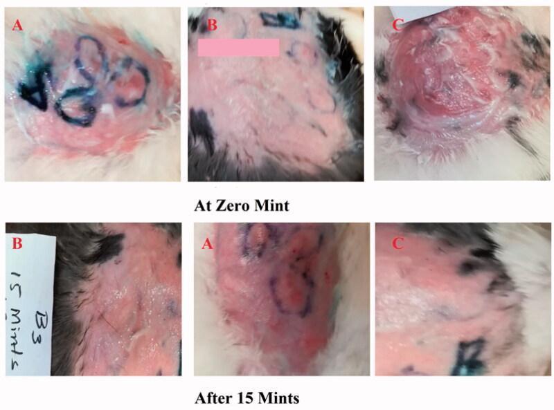
This comparison clinical photograph depicts the efficacy of topical antihistamine formulations on induced skin reactions in a rabbit model. The visual is organized into two rows representing time points: 'At Zero Mint' and 'After 15 Mints', subdivided into three experimental groups (A, B, and C). Group A evaluates a novel Ebastine emulgel, Group B serves as a positive control using a standard antihistamine (Benadryl), and Group C acts as the untreated negative control. At 0 minutes, all groups show histamine-induced erythema and wheal formation, with Group C exhibiting the most severe, raised, and inflamed lesion. Surface markings in blue and pink demarcate treatment zones. At 15 minutes, Group A and Group B demonstrate a visible reduction in the intensity and area of erythema, indicating effective drug absorption and inhibition of the H1-receptor-mediated allergic response. In contrast, Group C maintains significant, persistent inflammation and redness. The images illustrate the clinical progression of allergic skin lesions and the therapeutic effect of topical emulgel formulations on inflammatory symptoms like redness and swelling.
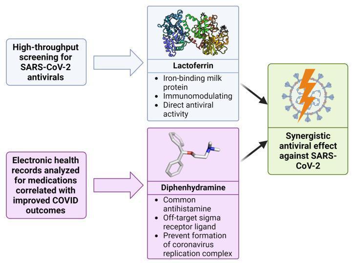
This medical flow diagram illustrates the pharmacological discovery and synergy between Lactoferrin and Diphenhydramine against SARS-CoV-2. The pathway starts with two screening methods: high-throughput screening for SARS-CoV-2 antivirals and electronic health record analysis for medications correlated with improved COVID outcomes. These lead respectively to Lactoferrin and Diphenhydramine. Lactoferrin is depicted as a complex structural protein model and described as an iron-binding milk protein with immunomodulating and direct antiviral activity. Diphenhydramine is shown as a chemical ball-and-stick model and identified as a common antihistamine and off-target sigma receptor ligand that prevents the formation of the coronavirus replication complex. Both agents point toward a final node represented by a SARS-CoV-2 virion with a lightning bolt symbol, signifying a 'Synergistic antiviral effect against SARS-CoV-2.' The diagram serves as an educational summary of how multi-modal screening can identify repurposed drug combinations that target different stages of the viral life cycle to enhance therapeutic efficacy.
Antihistamines
Overview
Histamine Receptors - A Primer
| Receptor | Coupling | Primary Actions |
|---|---|---|
| H1 | G-protein → phospholipase C | Smooth muscle contraction, vasodilation, increased capillary permeability, itching, pain, bronchoconstriction |
| H2 | G-protein → adenylyl cyclase | Gastric acid secretion (primary), some cardiac chronotropy |
| H3 | Presynaptic autoreceptor | Modulates CNS neurotransmitter release |
| H4 | Immune cells | Chemotaxis, immune modulation |
H1 Receptor Antagonists
Chemistry and Mechanism
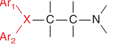
Generations of H1 Antihistamines
First-Generation (Sedating)
- Lipophilic - readily cross the blood-brain barrier
- Block both peripheral H1 and CNS histamine receptors
- Also bind muscarinic (anticholinergic) and adrenergic receptors
- Cause significant sedation - used therapeutically as sleep aids and for motion sickness
- Classified into chemical subclasses:
- Ethanolamines (diphenhydramine)
- Alkylamines (chlorpheniramine)
- Piperazines (hydroxyzine, meclizine, cyclizine)
- Piperidines (cyproheptadine)
- Phenothiazines (promethazine)
- Ethylenediamines (pyrilamine)
Second-Generation (Non-sedating)
- Highly specific for peripheral H1 receptors
- Do not penetrate the CNS (or do so minimally) - minimal sedative/anticholinergic effects
- Longer half-lives - most are once-daily dosing
- Fexofenadine is considered the least sedating of all
Pharmacological Effects of H1 Antagonists
| System | Effect |
|---|---|
| Smooth muscle | Inhibit histamine-induced bronchoconstriction and gut contraction |
| Vasculature | Reduce increased capillary permeability, attenuate wheal-and-flare response |
| Exocrine glands | Reduce nasal/bronchial secretions (1st gen more than 2nd gen) |
| CNS (1st gen) | Sedation, antiemetic, antiparkinsonian, local anesthetic at high doses |
| Skin | Relieve urticaria and pruritus |
Clinical Uses
H1 Antagonists
- Allergic rhinitis - first-line; 2nd-gen preferred for daytime use
- Urticaria (acute and chronic) - most striking benefit; chronic urticaria may need doses up to 4x the rhinitis dose
- Pruritus - atopic/contact dermatitis, insect bites, poison ivy
- Conjunctivitis - allergic eye symptoms
- Motion sickness - dimenhydrinate, meclizine, promethazine (scopolamine is superior)
- Vertigo / Ménière disease - dimenhydrinate, meclizine
- Insomnia - diphenhydramine (OTC sleep aids)
- Nausea/vomiting - promethazine (chemo-induced); 5-HT3 antagonists are more effective
- Extrapyramidal reactions - diphenhydramine reverses antipsychotic-induced dystonia
- Serum sickness urticaria - H1 antagonists help; fever and arthralgia often do not respond
- Anaphylaxis - adjuvant role only; epinephrine is the mainstay
In bronchial asthma, H1 antihistamines have limited efficacy and are not used as sole therapy. (Goodman & Gilman's)
Adverse Effects
First-Generation H1 Antagonists
- Sedation - most common; additive with alcohol and other CNS depressants
- Dizziness, tinnitus, lassitude, incoordination
- Anticholinergic effects: dry mouth, urinary retention, constipation, blurred vision, tachycardia
- In young children and at toxic doses: CNS excitation, restlessness, hallucinations, seizures
- Overdose: CNS depression or stimulation + peripheral anticholinergic effects + seizures
- QT prolongation (especially with 1st-gen and some 2nd-gen agents)
Second-Generation H1 Antagonists
- Generally well tolerated
- Cetirizine has mild sedation at therapeutic doses
- Rare QT prolongation (terfenadine and astemizole - both withdrawn from market due to this)
- Fexofenadine and loratadine considered safest cardiac profile
Toxicology Notes
- High concentrations of diphenhydramine and promethazine can cause false positives on urine immunoassay screens for amphetamines, TCAs, and propoxyphene
- Antihistamines have been used in drug-facilitated sexual assault (detected by forensic labs)
- Quantitative serum levels correlate poorly with clinical effects and are rarely clinically useful
H2 Receptor Antagonists
- Block H2 receptors on gastric parietal cells → reduce gastric acid secretion
- Uses: peptic ulcer disease, GERD, Zollinger-Ellison syndrome, stress ulcer prophylaxis
- Cimetidine is a potent CYP450 inhibitor (multiple drug interactions); others are less problematic
- Ranitidine was withdrawn globally (2019-2020) due to NDMA (carcinogen) contamination concerns
Special Populations
| Population | Consideration |
|---|---|
| Pregnancy | Chlorpheniramine, loratadine generally preferred; promethazine used for hyperemesis |
| Elderly | Avoid 1st-gen (Beers Criteria) - high anticholinergic burden, fall risk, cognitive impairment |
| Children | 1st-gen cause paradoxical excitation; 2nd-gen preferred for >2 years |
| Urticaria refractory to H1 blockers | Add H2 antagonist (H1 + H2 combination) + montelukast |
Key Comparison: 1st vs 2nd Generation
| Feature | 1st Generation | 2nd Generation |
|---|---|---|
| CNS penetration | High | Low/None |
| Sedation | Significant | Minimal |
| Anticholinergic | Yes | No |
| Dosing frequency | 3-4x daily | Once daily |
| Duration of action | 4-6 hours | 12-24 hours |
| Example | Diphenhydramine | Fexofenadine |
- Goodman & Gilman's Pharmacological Basis of Therapeutics - Chapter 43 (Histamine, Bradykinin, and Their Antagonists)
- Tietz Textbook of Laboratory Medicine, 7th Ed. - Antihistamines section
- Goldman-Cecil Medicine - Antipruritic Agents
- Harrison's Principles of Internal Medicine 22E - Urticaria and Angioedema