
Lymph node metastasis in unknown primary in detail according to standard ENT textbook in simplified manner
I'll start by reading the medical library skill, then search for relevant content on lymph node metastasis with unknown primary.
Reading File
Now let me search the library in parallel for all relevant content on this topic.
Finding Sources
Finding Sources
Finding Sources
Good - found content in Scott-Brown's and Cummings. Let me now find the dedicated sections on unknown primary in both textbooks.
Finding Sources
Finding Sources
Excellent! Found the key sections. Let me read all three major sources in parallel.
Reading File
Finding Sources
Reading File
Now let me read the Cummings dedicated "UNKNOWN PRIMARY" section and also the main Scott-Brown chapter on CUP.
Reading File
Reading File
Your credits are exhausted. You can purchase a top-up or subscribe to a plan to continue using Orris. If you're already subscribed, your daily credits will reset tomorrow.
Lymph node metastasis in unknown primary in detail according to standard ENT textbook in simplified manner
Reading File
Finding Sources
Finding Sources
Excellent - found all key ENT sources. Now reading all major sections in parallel.
Reading File
Reading File
Reading File
Good content retrieved. Now I need the detailed CUP chapter from Scott-Brown's main volume and the Cummings block 4288 section.
Reading File
Reading File
Now let me get the detailed CUP chapter from Scott-Brown's covering definitions, workup, nodal levels, and treatment.
Reading File
Excellent content. Let me now get the earlier part of this chapter for definition, epidemiology, and node-level correlations.
Reading File
Reading File
Now let me fetch the algorithm image and search for relevant images, while also getting the remaining radiotherapy and prognosis sections.
Loading Image
Reading File
I now have comprehensive content from multiple standard ENT textbooks. Let me compile the full, detailed answer.
Lymph Node Metastasis from Unknown Primary (CUP) - Head & Neck
Based on Scott-Brown's Otorhinolaryngology Head & Neck Surgery and Cummings Otolaryngology Head & Neck Surgery
1. What is CUP?
Carcinoma of Unknown Primary (CUP) is when a patient presents with biopsy-proven squamous cell carcinoma (SCC) in one or more cervical lymph nodes, but no obvious primary tumour is found despite:
- Thorough clinical examination
- Cross-sectional imaging (CT/MRI)
- Examination under anaesthesia (EUA) with tonsillectomy and tongue base biopsy
True CUP (strict definition): Metastatic neck lymphadenopathy where no primary tumour develops or is found within a subsequent 5-year follow-up period.
CUP accounts for up to 5% of all head and neck malignancies, but this figure is shrinking as detection techniques improve. However, the absolute number may be increasing due to the rising incidence of HPV-positive oropharyngeal cancers.
2. Why Does CUP Happen?
Two main mechanisms explain CUP:
| Mechanism | Explanation |
|---|---|
| Small primary + fast nodal growth | A tiny primary tumour metastasizes early; the cervical node grows much faster than the primary, which remains clinically invisible |
| Immune regression | The primary site is destroyed by the host immune response while the metastasis persists |
Historical note: In 1882, Volkmann believed CUP arose from degeneration of a branchial cyst into carcinoma ("branchiogenic carcinoma") - but this theory has been largely abandoned. Martin et al. in 1950 set strict criteria to diagnose true branchiogenic carcinoma, which very few cases actually meet.
3. Most Common Likely Primary Sites
The oropharynx is the most common site harbouring an occult primary. The key subsites:
- Palatine tonsil - single most common occult primary site
- Tongue base (lingual tonsil)
- Nasopharynx
- Pyriform sinus
- Infrahyoid epiglottis
4. Nodal Level and Likely Primary Site Correlation
The level of the involved lymph node gives a strong clue about where the primary might be hiding:
| Nodal Level | Most Likely Primary Site |
|---|---|
| Level I | Oral cavity, lip, floor of mouth (almost NEVER nasopharynx) |
| Level II | Most common level in CUP; oropharynx, nasopharynx, oral cavity |
| Level III | Oropharynx, hypopharynx, larynx |
| Level IV | Hypopharynx, larynx, cervical oesophagus, thyroid |
| Level V | Nasopharynx, skin of posterior scalp |
Levels I-IV involvement is almost exclusively associated with upper aerodigestive tract SCC, with Level II being the most common.
5. Role of HPV and EBV
Two viral markers dramatically change the diagnostic approach:
HPV (Human Papillomavirus)
- p16 IHC positivity in the metastatic node reliably predicts oropharyngeal origin (tonsil or tongue base)
- Typical patient: middle-aged man, minimal smoking/alcohol history, cystic neck mass
- Often misdiagnosed as branchial cleft cyst - all lateral cystic neck masses in patients over 35 must be considered malignant until proven otherwise
- p16 positivity → perform bilateral tonsillectomy + lingual tonsillectomy (diagnostic AND therapeutic)
EBV (Epstein-Barr Virus)
- EBV serology positivity → strongly indicates nasopharyngeal primary
- EBV is the only tumour marker of clinical value in CUP
Under the current TNM (8th edition) staging: T0 category is only retained for HPV-associated oropharyngeal SCC and nasopharyngeal carcinoma (where viral testing can confirm the primary origin). For all other CUP cases, T0 is eliminated.
6. Workup / Investigation Algorithm
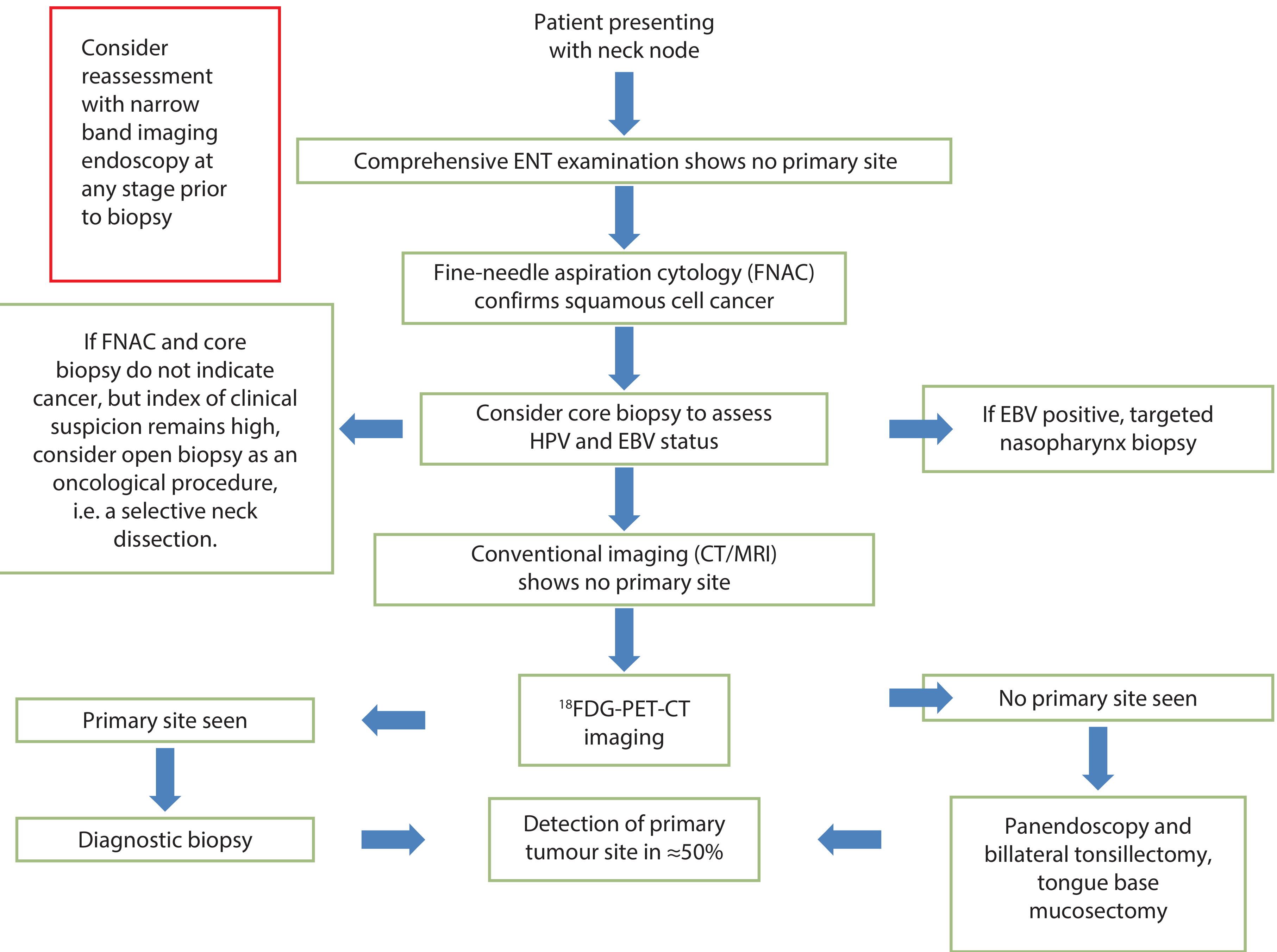
Step-by-step diagnostic approach:
Step 1 - History and Clinical Examination
- Heavy smoking/alcohol → primary outside nasopharynx
- Multiple sexual partners / orogenital contact → oropharyngeal primary (HPV-related)
- Fibreoptic nasolaryngoscopy with special attention to tongue base, nasopharynx, infrahyoid epiglottis, pyriform sinus
Step 2 - FNAC (First)
- Always get FNAC before imaging (to avoid PET-CT artefacts from prior biopsy)
- Confirms SCC histology
- Can test HPV (ISH + p16 IHC) and EBV on the aspirated material
Step 3 - Core Biopsy
- To assess HPV and EBV status if FNAC is non-diagnostic
Step 4 - Cross-sectional Imaging (CT and/or MRI)
- Identifies the primary in many cases
- MRI with diffusion sequences is increasingly useful
Step 5 - FDG PET-CT (KEY investigation)
- Must be done before any biopsy/EUA to avoid false positives
- NICE recommends considering PET-CT as the first imaging investigation
- Meta-analysis results:
- FDG PET-CT: detects 31.4% more primary sites than conventional workup alone
- Sensitivity 0.89, Specificity 0.73
- Also detects occult distant metastases and synchronous cancers (lung, colorectal)
- If EBV positive on biopsy → targeted nasopharynx biopsy
Step 6 - EUA + Panendoscopy + Surgical Biopsies
Performed if PET-CT still shows no primary:
- Panendoscopy (rigid pharyngolaryngoscopy)
- Bilateral tonsillectomy
- Tongue base mucosectomy (lingual tonsillectomy)
- Directed biopsies of nasopharynx, pyriform sinus, and any suspicious area
- Consider Narrow Band Imaging (NBI) endoscopy to highlight subtle mucosal abnormalities
7. Modern Surgical Approaches to Find the Primary - TORS / TLM
Traditional panendoscopy found the primary in only 17-40% of cases. Newer transoral approaches have transformed this:
Transoral Laser Microsurgery (TLM)
Using a CO2 laser and operating microscope, the surgeon:
- Inspects all mucosal surfaces under high magnification
- Looks for subtle signs: pallor, neovascularity, corkscrew telangiectasia, papillary growth, mucosal friability
- Takes a "cut-surface view" biopsy - the laser cut surface of tumour is paler and drier than surrounding tissue
- Sends to frozen section; if positive → resects to negative margins
Results with TLM:
| Approach | Primary Detection Rate |
|---|---|
| Traditional panendoscopy alone | 17-40% |
| TLM-assisted EUA (Karni et al.) | 94% |
| TLM-assisted (Nagel et al.) | 86.1% |
Transoral Robotic Surgery (TORS)
- Similar superior results to TLM
- Palatine + lingual tonsillectomy performed robotically
- Particularly powerful in p16-positive cases (89% detection rate in one series)
Key principle: If p16 IHC is positive on the neck node aspirate and nasopharyngoscopy is clear → go straight to transoral palatine + lingual tonsillectomy (it diagnoses AND treats the two most likely sites, avoiding wide-field pharyngeal radiotherapy)
8. Treatment
CUP management is divided into early (N1, no extracapsular spread) vs advanced (N2-N3 or extracapsular spread) disease:
ENT-UK MDT Treatment Guidelines Summary:
| Stage | Surgery | Radiotherapy | Chemotherapy |
|---|---|---|---|
| T0N1M0 (no ECS) | SND or MRND | Not routinely (consider for mucosal sites) | No |
| T0N1M0 (with ECS) | SND or MRND | Yes, to neck | Should be considered |
| T0N2-3M0 (no ECS) | SND or MRND | Yes, neck ± mucosal | Consider |
| T0N2-3M0 (with ECS) | SND or MRND | Yes, neck + mucosal | Yes (CRT) |
Surgical Management of the Neck
- N1 (no ECS): Selective neck dissection (SND) or surveillance alone after RT
- N2a-N2b: Selective neck dissection is valid (risk of level I and V metastasis is rare in CUP unless N3)
- N3 or ECS: MRND + adjuvant CRT
- "Violated neck" (prior open biopsy): Not a contraindication to good outcomes with current multimodal management
Radiation Therapy Controversy
Two schools of thought:
| Approach | Advantage | Disadvantage |
|---|---|---|
| Total mucosal irradiation (bilateral neck + all mucosa) | Better local control, lower primary site recurrence | High xerostomia, mucositis, dysphagia rates |
| Selective mucosal irradiation (ipsilateral, exclude nasopharynx) | Lower morbidity | Slightly higher risk of contralateral/distant recurrence |
- IMRT is the current standard - allows parotid gland sparing and significantly reduces xerostomia
- Total mucosal irradiation dose: 50 Gy in 25 daily fractions is adequate for occult disease control
- Nodal stage is the most important risk factor for local recurrence
9. Prognosis and Patterns of Failure
Survival:
- Overall 5-year survival for all stages: 52-75%
- N1 disease: up to 100% 5-year survival
- N2-N3 disease: ~60% 5-year survival
- Recent series from Memorial Sloan Kettering (IMRT): 2-year regional progression-free survival 90%, OS 85%
Patterns of Failure:
- Primary site emergence: 0-66% (mainly in patients treated with surgery alone); usually occurs within 24 months, most often in the oral cavity, oropharynx, and nasopharynx
- Distant metastases: Occur within 1 year of treatment completion; lung is the most common site
- FDG PET-CT is the best method for detecting recurrence
10. Key Summary Points (Quick Recall)
| Point | Detail |
|---|---|
| Definition | SCC in cervical node, no primary found for 5 years |
| Incidence | <5% of head and neck malignancies |
| Most common primary site | Oropharynx (tonsil > tongue base) |
| Most common nodal level | Level II |
| Most important tumour marker | EBV (→ nasopharynx); HPV/p16 (→ oropharynx) |
| First-line biopsy | FNAC (before imaging) |
| Key imaging | FDG PET-CT (before EUA) |
| Best surgical detection | TLM / TORS-assisted EUA (~90%) |
| Lateral cystic neck mass >35 yr | Malignant until proven otherwise |
| Standard RT technique | IMRT |
| Best prognosis | N1, no ECS |
Sources: Scott-Brown's Otorhinolaryngology Head & Neck Surgery (Chapter 17 - CUP, Chapter 47 - Occult Primary Tumours); Cummings Otolaryngology Head & Neck Surgery (Chapter 96 - Unknown Primary; Chapter Metastatic Lesions)
TORS in detail according to standard ent textbook and recent modalities in simplified manner
Finding Sources
Finding Sources
Searching the Web
TORS transoral robotic surgery recent advances 2024 2025 ENT head neck
Excellent sources found. Now reading all the major TORS textbook sections in parallel.
Reading File
Reading File
Reading File
Great content flowing in. Let me now read the detailed Scott-Brown's Chapter 29 on TORS (indications, pre-op, technique, outcomes, TLM comparison) and the parapharyngeal space TORS section.
Reading File
Reading File
Now let me get the key remaining sections - TORS vs TLM comparison, outcomes, and Future Horizons from Cummings, plus fetch the da Vinci image.
Reading File
Reading File
Loading Image
I now have comprehensive textbook content. Let me also get the indications, contraindications, and outcomes data from the main Scott-Brown's TORS chapter.
Reading File
Searching PubMed
"transoral robotic surgery" AND oropharyngeal cancer outcomes
I now have all the content needed for a thorough, well-structured answer. Let me compile it.
Transoral Robotic Surgery (TORS) - Detailed ENT Textbook Review
Based on Scott-Brown's Otorhinolaryngology Head & Neck Surgery and Cummings Otolaryngology Head & Neck Surgery
1. What is TORS?
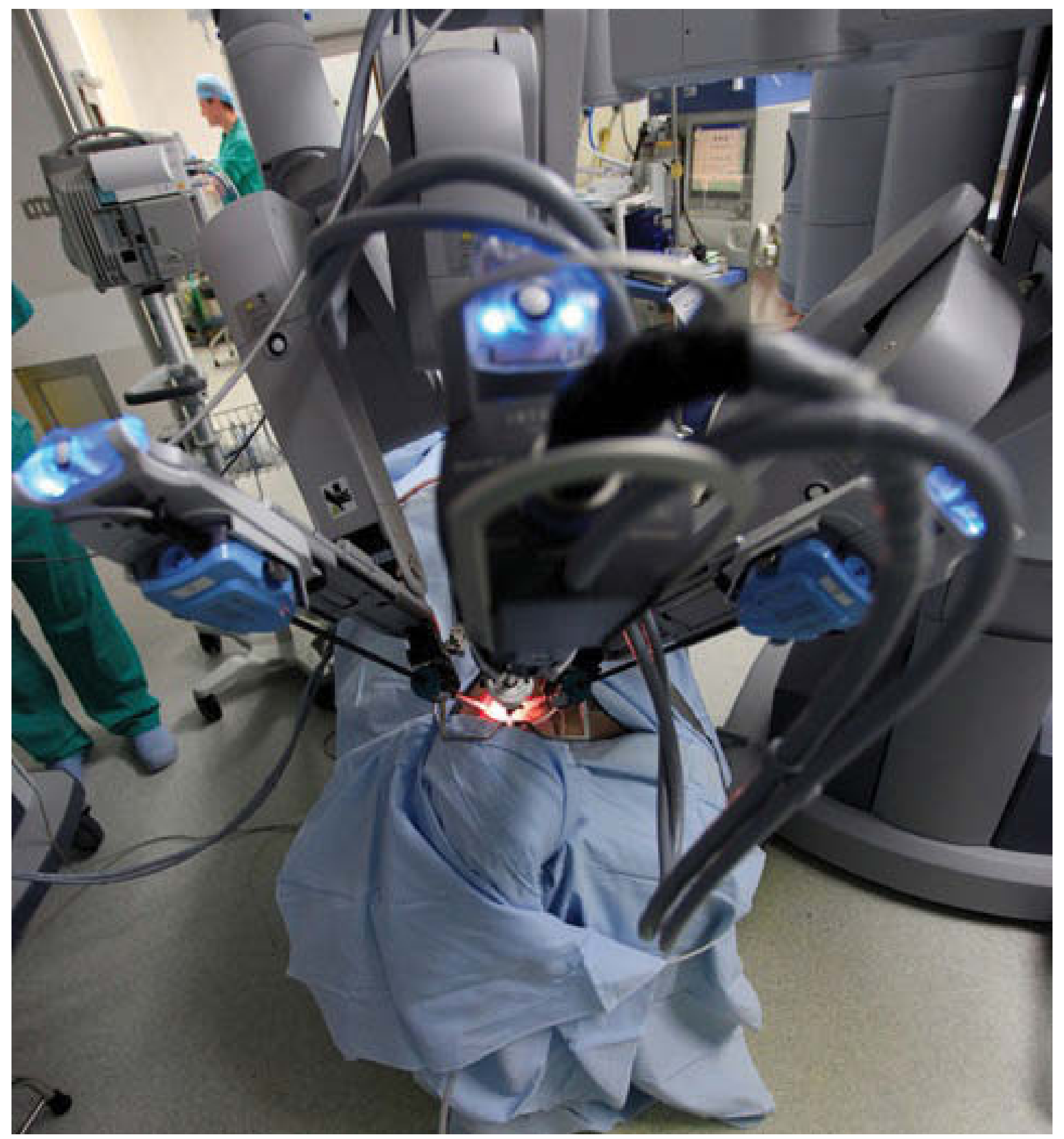
Transoral Robotic Surgery (TORS) is a minimally invasive surgical technique in which the surgeon operates on tumours of the oral cavity, pharynx, and larynx through the mouth, without any external skin incision, using a robotic surgical system (most commonly the da Vinci® system by Intuitive Surgical).
The surgeon controls miniaturised articulated instruments and a high-definition 3D camera from a console away from the patient, achieving precision, stability, and visualization that is impossible with conventional transoral instruments.
2. Historical Background
| Year | Milestone |
|---|---|
| 2003 | Melder & McLeod - first robotic procedure in otolaryngology (vallecular cyst removal at Walter Reed Army Medical Center) |
| 2005-2006 | Hockstein, Weinstein, and O'Malley publish landmark papers defining TORS using oral retractors (FK, Crowe-Davis, McIvor, Dingman) instead of laryngoscopes |
| 2006 | O'Malley et al. demonstrate TORS in cadaver/animal models then first 3 human tongue base cancers |
| 2007 | Weinstein et al. - radical tonsillectomy via TORS |
| 2009 | FDA approval for TORS for T1-T2 oropharyngeal tumours |
| 2026 | Celebrating 20 years of TORS - now used globally for oropharyngeal, supraglottic, hypopharyngeal, parapharyngeal, and thyroid surgery |
The key insight by Hockstein et al. was that oral and oropharyngeal retractors (rather than tubed laryngoscopes) provided relatively free motion of robotic arms for oropharynx, supraglottis and hypopharynx procedures.
3. The Robotic System Components
The da Vinci system has 4 main components:
| Component | Function |
|---|---|
| 1. Surgeon Console | Monitor + hand/foot controls; surgeon sits here and operates |
| 2. Patient-Side Cart | 3 robotic arms - 1 central arm (camera) + 2 lateral arms (instruments) |
| 3. Vision Cart | Displays the surgical field from the 0° or 30° endoscope |
| 4. EndoWrist Instruments | 7 degrees of freedom - far exceeds human wrist movement |
Key Instruments:
- 5 mm or 8 mm diameter interchangeable instruments
- Atraumatic forceps, monopolar cautery spatula (most common cutting/dissecting tool), bipolar cautery, dissectors
- 8.5 or 12 mm binocular stereoendoscopy camera - provides high-definition 3D visualization
- Fibre laser technology - now adaptable to robotic arms; more precise cutting, less thermal damage than monopolar cautery
Access to the Oropharynx:
Achieved with spatulate retractors:
- Feyh-Kastenbauer (FK) retractor - most common
- Crowe-Davis retractor
- McIvor retractor
- Dingman retractor
An assistant surgeon sits at the patient's side for suction and haemostasis.
4. Indications for TORS
FDA-Approved:
- T1-T2 oropharyngeal tumours (palatine tonsil, tongue base/BOT)
Commonly Used For:
- Oropharyngeal SCC - most common application; tonsil and tongue base tumours
- Supraglottic laryngeal cancer - supraglottic partial laryngectomy
- Unknown primary - bilateral tonsillectomy + lingual tonsillectomy (diagnostic + therapeutic)
- Parapharyngeal space tumours - benign salivary gland tumours, schwannomas, branchial cysts
- Hypopharyngeal tumours - selected cases
- Tongue base reduction for obstructive sleep apnoea (OSA)
- Benign conditions - epiglottoplasty, vallecular cysts, Eagle's syndrome, posterior glottic pathology
5. Contraindications
Absolute Contraindications (for oropharyngeal cancer):
- Inadequate transoral access / difficult mouth opening / trismus
- Incomplete tumour visualization despite spatulate retraction
- Deep extrinsic muscle invasion in tongue base tumours
- Invasion lateral to constrictor muscles into deep tissues
- Pre-vertebral fascia invasion
- Laryngeal involvement
- Retropharyngeal position of internal carotid artery (scans must be carefully studied)
- Tumour crossing the midline (BOT tumours)
For Parapharyngeal Space:
- Tumour adherent to or involving the carotid artery
- Vascular tumours (paragangliomas)
- Skull base bony involvement
- Malignant tumours of the parapharyngeal space
- Dumbbell tumours with significant parotid gland involvement
For Tonsil Tumours:
- Extension to parapharyngeal space and pterygoid musculature (relative contraindication)
6. Pre-operative Assessment
Before TORS, always assess:
- Mouth opening and neck mobility - adequate access is essential
- Cross-sectional imaging (CT/MRI) - assess tumour extent, confirm resectability, look for retropharyngeal carotid artery
- PET-CT - staging, identify unknowns
- HPV/p16 + EBV status - from FNAC of neck node
- Dental review - for radiation planning if adjuvant RT planned
- Anaesthetic assessment - airway, tracheostomy planning
7. Surgical Technique
General Setup:
- Patient supine, neck extended, mouth propped with retractor
- Robot docked over the patient's head
- Surgeon at console (may be in same room or remote)
- Assistant at bedside with suction + haemostasis
For Tonsil Tumours (Radical Tonsillectomy):
- Neck dissection performed first (same GA or prior sitting) - ipsilateral lingual and facial arteries are identified and ligated in continuity
- Resection by monopolar cautery (laser if available)
- En bloc removal - resection specimen is essentially a lateral oropharyngectomy including the constrictor muscle of the tonsil bed
- Margins checked intraoperatively - areas of concern marked with methylene blue for pathologist
- Further margins harvested if needed, especially at the inferior aspect where tonsil meets tongue base
- Specimen orientated on a mount and sent for histology
For Tongue Base (BOT) Tumours:
- Resectability confirmed on imaging
- Midline crossing is a contraindication
- Ipsilateral tongue base resection; further margins especially at deeper aspect of BOT
- Specimen orientated and sent for histology
For Tumours Involving Both Tonsil and BOT:
- Cut through tonsil and constrictors ≥5 mm from tumour edge
- Leave cranial part of tonsil, resect tonsil tumour to styloglossus
- Then make BOT cuts and remove entire specimen en bloc
Principle:
TORS = En bloc resection with clear margins (unlike TLM which uses the "cut-through" piecemeal technique)
8. TORS vs. Transoral Laser Microsurgery (TLM) - Key Comparison
| Feature | TORS | TLM |
|---|---|---|
| Resection principle | En bloc with clear margins | Piecemeal; cuts through tumour to follow extent |
| Visualization | HD 3D, magnified, tremor-filtered | 2D operating microscope, line-of-sight only |
| Margin reporting | Clear margins more easily achieved | Margins frequently reported as "close"; relies on marginal biopsies |
| Adjuvant planning | More definitive margin data → easier to plan | Marginal close results → uncertainty about adjuvant RT |
| Learning curve | Not as steep as TLM (superior optics + ergonomics help) | Steeper learning curve |
| Access | Better manoeuvrability (EndoWrist) | Limited by line-of-sight |
| En bloc tonsil | Easily performed | Technically difficult (line of sight limits it) |
| Primary detection (CUP) | ~89-94% (with mucosectomy) | ~86-94% |
| Cost | Higher | Lower |
The significant TLM literature paved the way for TORS to become established as a valid management of tonsil tumours with firm long-term oncological outcomes.
9. Outcomes Data
Safety (Weinstein et al., multicentre prospective - 177 patients):
- No intra-operative mortality
- Average blood loss: 83 mL (no transfusions)
- Serious adverse events: 16% (required intervention)
- Tracheostomy at follow-up: 2.3%
- Gastrostomy tube dependence: 5%
- Positive margin rate: 4.3%
Survival (White et al., 89 patients, 2 institutions):
- 2-year recurrence-free survival: 86.5%
- None of the patients were gastrostomy-tube dependent at follow-up
Moore et al. (45 patients, T1-T4a, prospective):
- All margins negative
- All tracheostomized patients eventually decannulated
- All feeding tube patients eventually had tubes removed
Recent Meta-Analyses (2024-2026):
- Gupta et al., 2024 (PMID 38795678) - Quality of life comparing TORS vs. primary radiotherapy: similar QOL outcomes, supporting TORS as a valid alternative
- Cheung et al., 2026 (PMID 41521853) - Oncologic outcomes in HPV-associated OPSCC: strong results confirming TORS efficacy in HPV+ disease
10. TORS for Specific Sites
A. Oropharyngeal Cancer (Most Common Application)
- Tonsil SCC and tongue base SCC (T1-T2, FDA-approved)
- HPV-positive OPSCC: TORS + selective neck dissection ± de-escalated adjuvant RT
- Goal: avoid or de-intensify chemoradiotherapy → reduce long-term xerostomia, dysphagia, feeding tube dependence
B. Supraglottic Larynx
- Supraglottic partial laryngectomy via TORS
- Avoids tracheostomy in many patients
- Functional preservation of voice and swallowing
C. Unknown Primary (CUP)
- Bilateral palatine tonsillectomy + lingual tonsillectomy
- Increases primary detection from 17-40% (traditional EUA) to 89-94%
- If p16 positive: TORS tonsil/tongue base surgery is both diagnostic AND therapeutic, eliminating the need for wide-field pharyngeal RT
D. Parapharyngeal Space
- Ideal candidates: Benign pre-styloid salivary gland tumours, schwannomas, branchial cysts
- Tumour must displace carotid posteriorly/laterally (not medially)
- Avoids the traditional transcervical or transparotid approach
- No external scar
E. Obstructive Sleep Apnoea (OSA)
- Tongue base reduction (lingual tonsillectomy) to reduce retrolingual obstruction
- First prospective study on single-port robotic tongue base resection for sleep apnoea reported 2025
11. Post-operative Care
- All patients managed in HDU overnight - for airway monitoring and oral bleeding observation
- Usually extubated after surgery; no routine need for prolonged intubation or tracheostomy
- Oral diet protocol:
- Day 0: Nil by mouth
- Day 1: Clear fluids
- Day 2: Free fluids
- Day 3+: Soft diet → normal diet
12. Complications
| Complication | Details |
|---|---|
| Post-operative haemorrhage | Most feared; raw oropharyngeal wound; can compromise airway |
| Dysphagia | Usually temporary; long-term rates much lower than open surgery |
| Tracheostomy | Required in minority; all should eventually be decannulated |
| Positive surgical margins | ~4.3% (better than historical open surgery rates) |
| Taste disturbance | From tongue base resection |
| Dental injury | From retractor placement |
| For parapharyngeal: | Vascular injury, lower cranial nerve injury (CN IX-XII), tumour spillage, "first bite syndrome" |
13. Ongoing Trials and Evidence Base
| Trial | Key Question |
|---|---|
| PATHOS (NCT02215265) | Can adjuvant RT be de-escalated (50 Gy vs 60 Gy vs none) in HPV+ve OPSCC after TORS? |
| CompARE (ISRCTN41478539) | TORS + selective ND vs definitive CRT vs induction chemo + CRT in intermediate/high-risk OPSCC |
| RTOG 1221 | Transoral resection (p16+ OPSCC) + risk-based adjuvant RT (60 Gy ± cisplatin) vs definitive CRT |
No randomized trials yet directly comparing TORS to chemoradiation as primary treatment, but multiple Phase II/III trials are underway.
14. Recent Advances and Future Horizons (2024-2026)
The field is rapidly evolving:
| Advance | Description |
|---|---|
| Single-Port (SP) da Vinci robot | One port, multiple instruments - reduces crowding of working space, better access for tongue base; growing evidence 2024-2026 |
| Laser-coupled robotic arms | CO2 fibre laser and Thulium:YAG (Tm:YAG) laser attached to robotic arms - better cutting, less thermal damage than monopolar cautery |
| Flexible robotic cameras | Greater angulation, improved visualization of difficult anatomy |
| Haptic feedback | Surgeon can "feel" tissue resistance - currently absent in da Vinci |
| Tissue fluorescence + targeted markers | Improved intraoperative margin analysis; identifies tumour boundaries in real time |
| Image-guided TORS | Surgical navigation overlaid on robotic view |
| TORS for OSA | Single-port robotic tongue base resection - first prospective data 2025 |
| TORS for benign glottic surgery | Novel application - posterior glottic scar lysis and partial arytenoidectomy (2025) |
| Versius system (CMR Surgical) | Alternative to da Vinci; growing use in TORS |
| New smaller robotic systems | Microsurgical robots with finer instruments being explored |
| De-escalation trials | PATHOS, RTOG 1221 - aiming to reduce RT toxicity in HPV+ patients treated with TORS |
15. Key Summary Points
| Point | Detail |
|---|---|
| What is TORS | Surgery through the mouth using a robotic system |
| Main platform | da Vinci® Si/Xi/SP systems |
| FDA approval | T1-T2 oropharyngeal cancer |
| Key advantage over TLM | En bloc resection, 3D HD visualization, better ergonomics |
| Key advantage over open | No external scar, less morbidity, faster recovery |
| Most common indication | Oropharyngeal SCC (tonsil + tongue base) |
| Best patient for CUP | HPV+ cystic neck node, p16 positive - tonsil/BOT surgery is diagnostic + therapeutic |
| Main cutting tool | Monopolar electrocautery (laser increasingly used) |
| Main retractor | Feyh-Kastenbauer (FK) |
| Key contraindication | Inadequate mouth opening, carotid involvement, midline BOT, pre-vertebral invasion |
| 2-year RFS | ~86.5% (early stage OPSCC) |
| Future | SP robot, laser-coupled arms, haptic feedback, tissue fluorescence, de-escalation trials |
Sources: Scott-Brown's Otorhinolaryngology Head & Neck Surgery - Chapters 17, 29, Chapter 11 (Parapharyngeal); Cummings Otolaryngology Head & Neck Surgery - Chapters 96, 97 (Future Horizons); Recent meta-analyses PMID 38795678, 41521853
This is a shared conversation. Sign in to Orris to start your own chat.