I now have everything I need from the textbooks. Let me compile the comprehensive answer.
Everything You Need to Know Before Reading Systemic Pharmacology
This is your complete foundational framework - receptor biology, pharmacodynamics, and pharmacokinetics - drawn from Katzung's Basic and Clinical Pharmacology, 16th Edition (Chapters 1 & 2).
1. What Is Pharmacology?
Pharmacology = the study of substances that interact with living systems through chemical processes, usually by binding to regulatory molecules and activating or inhibiting normal body processes.
Two major branches:
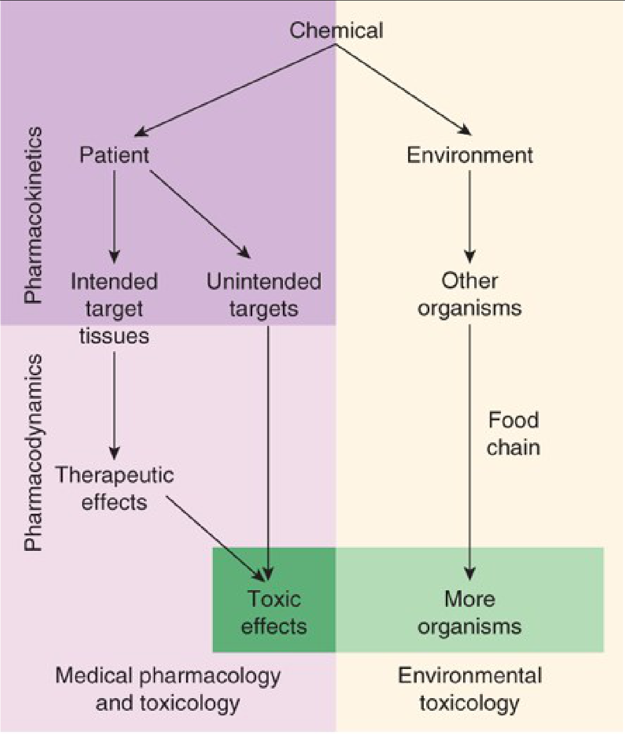
- Pharmacokinetics - what the body does to the drug (absorption, distribution, metabolism, elimination)
- Pharmacodynamics - what the drug does to the body (receptor binding, mechanism, effect)
2. Drug Properties
Size
- Most drugs: MW 100-1000
- Below 100 MW = too small to bind with specificity
- Above 1000 MW = too large to diffuse between body compartments (must be given IV or directly at site)
- Large biologics (antibodies, proteins) often >100,000 MW
Drug-Receptor Bond Types
Three types of bonds form between drugs and receptors:
| Bond Type | Strength | Reversibility | Example |
|---|
| Covalent | Very strong | Usually irreversible | Aspirin + COX (platelet); DNA alkylating agents |
| Electrostatic | Moderate | Reversible | Most drug-receptor interactions (ionic, hydrogen bonds, van der Waals) |
| Hydrophobic | Weak | Reversible | Lipid-soluble drugs in membrane "pockets" |
Key insight: Weak bonds = higher selectivity - the drug must fit its receptor very precisely. Strong covalent bonds are less selective.
Drug Shape
Shape must be complementary to the receptor (like a key fitting a lock). Chirality matters - most drugs are racemic mixtures, but the two enantiomers can have very different receptor affinities, potencies, and even different effects.
Ionization (Henderson-Hasselbalch)
- Most drugs are weak acids or bases
- Ionized form = water-soluble, cannot cross lipid membranes
- Un-ionized form = lipid-soluble, crosses membranes freely
Henderson-Hasselbalch equation:
pH = pKa + log([ionized]/[un-ionized]) for bases
For a weak acid (like aspirin, pKa 3.5):
- In stomach (pH 1-2): mostly un-ionized → absorbed
- In blood (pH 7.4): mostly ionized → trapped
Ion trapping: Alkalinizing the urine (sodium bicarbonate) ionizes weak acid drugs → traps them in urine → increases renal elimination (this is why NaHCO₃ is given in aspirin overdose).
3. Drug Permeation Across Membranes
Four mechanisms:
- Aqueous diffusion - through water-filled channels (pores) in endothelium; small polar molecules
- Lipid diffusion - the most important for most drugs; depends on lipid:water partition coefficient
- Special carriers (transporters) - active or facilitated transport; for molecules too polar to diffuse (eg, glucose, amino acids, organic acids)
- Endocytosis/exocytosis - for very large molecules (eg, vitamin B12-intrinsic factor complex, iron-transferrin)
Fick's Law of Diffusion:
Flux = (Area × Permeability coefficient × [C1-C2]) / Thickness
4. The Receptor Concept - Core Foundation
A receptor is the macromolecule in a cell or organism that interacts with a drug and initiates the chain of events leading to the drug's observed effects.
Receptors serve two fundamental functions:
- Ligand binding - recognition and binding of the drug/signal
- Signal transduction - converting binding into a cellular response
Receptors are primarily proteins - cell surface or intracellular. Some important drug targets are not traditional "receptors" but enzymes (aspirin/COX), ion channels (local anesthetics/Na channels), transport proteins (SSRIs/serotonin transporter), or structural proteins.
5. The Four Major Receptor Families
Type 1: Ligand-Gated Ion Channels (Ionotropic Receptors)
- Structure: Receptor IS the ion channel - ligand binds and directly opens/closes the channel
- Speed: Milliseconds (fastest)
- Location: Cell surface
- Mechanism: Conformational change opens pore → ion flow → membrane potential change
- Examples:
| Receptor | Ion | Effect |
|---|
| Nicotinic ACh (nAChR) | Na⁺ in / K⁺ out | Depolarization (excitatory) |
| GABA-A | Cl⁻ in | Hyperpolarization (inhibitory) |
| Glycine | Cl⁻ in | Hyperpolarization (inhibitory) |
| NMDA (glutamate) | Na⁺, Ca²⁺ in | Excitatory, LTP |
| AMPA (glutamate) | Na⁺, K⁺ | Fast excitatory |
| 5-HT3 (serotonin) | Na⁺, K⁺ | Excitatory |
- Drug targets: Benzodiazepines (modulate GABA-A), succinylcholine/d-tubocurarine (nAChR), memantine (NMDA)
Type 2: G Protein-Coupled Receptors (GPCRs) / Metabotropic
- Structure: 7 transmembrane (7-TM) helical domains; largest receptor superfamily in the human genome (~800 genes)
- Speed: Seconds to minutes
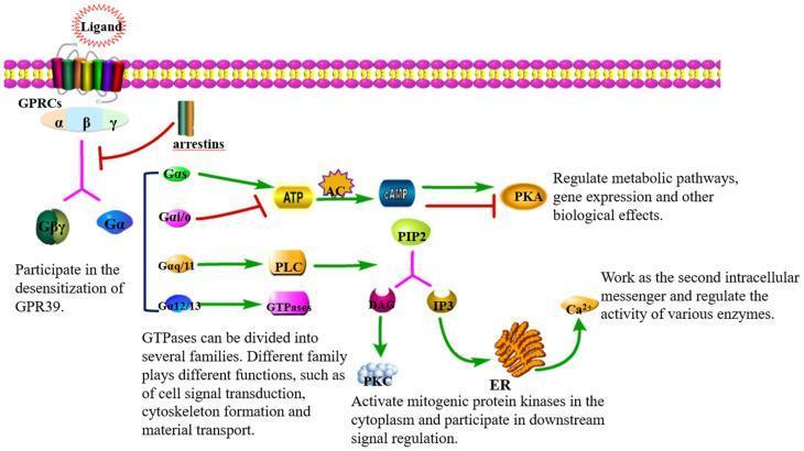
- Mechanism: Ligand binds → activates G protein (trimeric: Gα, Gβ, Gγ) → Gα dissociates → acts on effectors (enzymes or ion channels)
G protein subtypes and their effectors:
| G Protein | Second Messenger / Effect | Examples |
|---|
| Gs | ↑ Adenylyl cyclase → ↑ cAMP → ↑ PKA | β1-AR (heart), β2-AR (bronchi), D1 receptor, H2 receptor |
| Gi | ↓ Adenylyl cyclase → ↓ cAMP | M2 muscarinic, D2, α2-AR, opioid (μ,δ,κ) |
| Gq/G11 | ↑ Phospholipase C → ↑ IP3 + DAG → ↑ Ca²⁺ + PKC | M1, M3, M5, α1-AR, H1, AT1 |
| G12/13 | Activates Rho GTPases → cytoskeleton | Thrombin, LPA receptors |
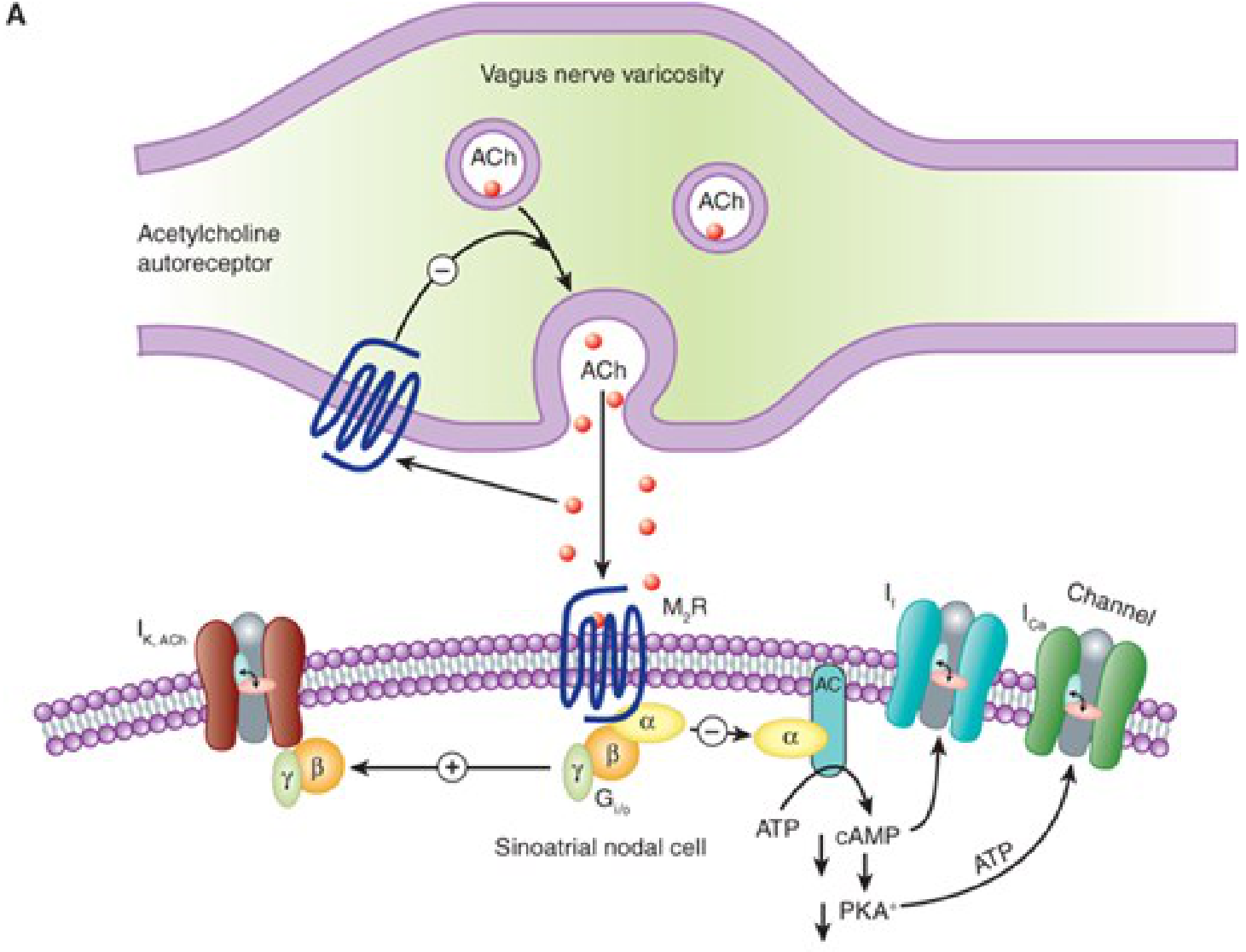
| Gβγ (free subunit) | Directly opens GIRK K⁺ channels | M2 → K⁺ channel → slows SA node |
Second messenger cascade:
- cAMP pathway: Receptor → Gs → Adenylyl cyclase → ATP → cAMP → PKA activation → phosphorylation of proteins (eg, L-type Ca²⁺ channels, glycogen phosphorylase)
- IP3/DAG pathway: Receptor → Gq → PLC-β → PIP2 → IP3 (releases ER Ca²⁺) + DAG (activates PKC)
Type 3: Enzyme-Linked Receptors (Receptor Tyrosine Kinases / RTKs)
- Structure: Single transmembrane domain; extracellular ligand-binding domain + intracellular kinase domain
- Speed: Minutes to hours
- Mechanism: Ligand binds → receptor dimerization → autophosphorylation on tyrosine residues → recruits adaptor proteins → activates downstream cascades (MAPK/ERK, PI3K/Akt, JAK/STAT)
- Examples:
| Receptor | Ligand | Signaling |
|---|
| Insulin receptor | Insulin | Tyrosine kinase → PI3K/Akt → GLUT4 translocation |
| EGF receptor | EGF | Tyrosine kinase → MAPK/ERK → cell growth |
| VEGF receptor | VEGF | Angiogenesis |
| Cytokine receptors (JAK-STAT) | Interferons, EPO, IL-6 | JAK phosphorylates STAT → gene transcription |
- Drug targets: Imatinib (BCR-ABL tyrosine kinase), trastuzumab (HER2), erlotinib (EGFR)
Type 4: Intracellular (Nuclear) Receptors
- Location: Cytoplasm or nucleus
- Ligands: Lipid-soluble small molecules that diffuse through the plasma membrane
- Speed: Hours to days (must synthesize new proteins)
- Mechanism: Ligand binds receptor → releases inhibitory protein (eg, hsp90 for glucocorticoid receptor) → receptor dimerizes → translocates to nucleus → binds DNA response elements → activates/represses gene transcription
Two-step mechanism for glucocorticoids:
- Steroid diffuses into cell
- Binds GR → hsp90 dissociates → GR dimerizes → enters nucleus → binds GRE (glucocorticoid response element) → mRNA synthesis
| Ligand | Receptor | Effect |
|---|
| Glucocorticoids | GR | Anti-inflammatory gene expression |
| Mineralocorticoids | MR | Sodium retention genes |
| Sex steroids | AR, ER, PR | Secondary sex characteristics, reproduction |
| Thyroid hormone (T3) | TR | Metabolic rate |
| Vitamin D | VDR | Calcium homeostasis genes |
| Retinoids | RAR, RXR | Differentiation, growth |
Clinical consequence: Glucocorticoids do NOT relieve asthma immediately - there is a lag of 30 min to several hours for new protein synthesis.
6. Receptor Regulation
Desensitization / Tachyphylaxis / Down-regulation:
- Prolonged agonist exposure → receptor phosphorylation by GRKs → β-arrestin binding → receptor internalization (endocytosis) → fewer surface receptors → reduced response
- Example: Prolonged β2-agonist use in asthma → tachyphylaxis
Supersensitivity / Up-regulation:
- Prolonged antagonist exposure → more receptors synthesized → exaggerated response if antagonist suddenly withdrawn
- Example: β-blocker withdrawal → rebound tachycardia/hypertension
7. Dose-Response Relationships
Graded Dose-Response Curves
- Plotted as effect (%) vs log[drug concentration]
- Sigmoidal shape
Key parameters:
- EC50 (ED50): Concentration producing 50% of maximal effect - measures potency
- Emax: Maximal effect achievable - measures efficacy
- Hill coefficient (n): Slope of the curve; indicates cooperativity
Potency ≠ Efficacy: A drug can be very potent (low EC50) but have low efficacy (low Emax), or vice versa. Clinically, efficacy matters more than potency.
Agonists vs Antagonists
| Term | Definition | Effect on curve |
|---|
| Full agonist | Produces maximal response at full receptor occupancy | Emax = 100% |
| Partial agonist | Intrinsic activity < 1; can't produce Emax even at 100% occupancy | Emax < 100% (can act as antagonist in presence of full agonist) |
| Inverse agonist | Binds receptor, produces opposite effect to agonist | Drives receptor to inactive state |
| Competitive antagonist | Binds same site as agonist, reversibly; shifts curve right | ↑ EC50, same Emax |
| Irreversible/non-competitive antagonist | Binds irreversibly or allosteric site; can't overcome with more drug | ↓ Emax |
| Physiologic antagonist | Two drugs with opposite physiologic effects (different receptors) | |
Spare receptors (receptor reserve): Maximum effect can occur when only a fraction of receptors are occupied. This means EC50 < Kd (the dissociation constant). Important for: understanding why partial agonists can block full agonists in tissues with spare receptors.
8. Quantal Dose-Response & Therapeutic Index
Quantal curves plot the frequency of an all-or-none response (eg, % of population that responds) vs log dose.
- ED50: Dose effective in 50% of population
- LD50: Dose lethal in 50% of population (animal data)
- TD50: Dose producing a toxic effect in 50% of population
Therapeutic Index (TI) = TD50 / ED50
- Higher TI = safer drug
- Example: Digoxin has a narrow TI (toxic dose close to therapeutic dose)
Certain Safety Factor = TD1 / ED99 (more clinically useful; avoids any toxicity)
9. Pharmacokinetics Essentials
ADME
-
Absorption - rate and extent drug enters systemic circulation
- Bioavailability (F): Fraction of dose reaching systemic circulation
- Affected by: first-pass metabolism, solubility, formulation
-
Distribution
- Volume of distribution (Vd): Apparent volume needed to contain total drug at same concentration as plasma
- Small Vd = stays in plasma (eg, warfarin); Large Vd = distributes widely into tissues (eg, chloroquine)
- Blood-brain barrier: only lipid-soluble, un-ionized drugs cross
- Protein binding (albumin, α1-acid glycoprotein): only FREE drug is active
-
Metabolism
- Phase I: CYP450 enzymes - oxidation, reduction, hydrolysis → add/expose functional groups
- Phase II: Conjugation (glucuronidation, sulfation, acetylation, methylation) → increase polarity for excretion
- First-pass effect: oral drug metabolized in gut wall and liver before reaching systemic circulation
-
Elimination
- Renal: Filter + secrete (active) + reabsorb; affected by pH, protein binding
- Hepatic: Biliary excretion; enterohepatic cycling
Key PK Parameters
- Half-life (t½) = 0.693 × Vd / CL (CL = clearance)
- After 4-5 half-lives: drug reaches steady-state (if dosed repeatedly) OR drug is 97% eliminated
- Clearance (CL): Volume of plasma cleared per unit time; most important PK parameter for dosing
- Zero-order kinetics: Fixed amount eliminated per unit time (saturable; eg, alcohol, phenytoin at high doses) - dangerous nonlinear behavior
- First-order kinetics: Fixed fraction eliminated per unit time (most drugs)
10. The Autonomic Nervous System - The Most-Tested Receptor System
Before systemic pharmacology, you must know the ANS cold:
Sympathetic (Adrenergic) Receptors
| Receptor | Location | G protein | Effect | Key Agonists/Antagonists |
|---|
| α1 | Vascular smooth muscle, bladder, eye | Gq → IP3/DAG → ↑Ca²⁺ | Vasoconstriction, mydriasis, urinary retention | Agonist: phenylephrine; Antagonist: prazosin, tamsulosin |
| α2 | Presynaptic (autoreceptor), pancreatic β-cells | Gi → ↓cAMP | ↓NE release (negative feedback), ↓insulin | Agonist: clonidine; Antagonist: yohimbine |
| β1 | Heart (SA node, AV node, ventricle) | Gs → ↑cAMP | ↑HR, ↑contractility, ↑conduction | Agonist: dobutamine; Antagonist: metoprolol |
| β2 | Bronchial smooth muscle, vascular SM, uterus | Gs → ↑cAMP | Bronchodilation, vasodilation, uterine relaxation | Agonist: salbutamol; Antagonist: propranolol (non-selective) |
| β3 | Adipose tissue | Gs → ↑cAMP | Lipolysis, thermogenesis | Agonist: mirabegron (bladder) |
| DA1 (dopamine) | Renal/mesenteric vasculature | Gs → ↑cAMP | Vasodilation (renal protective) | Agonist: low-dose dopamine |
| DA2 | Presynaptic (autoreceptor) | Gi | ↓NE/DA release | |
Parasympathetic (Cholinergic) Receptors
Muscarinic Receptors (GPCRs):
| Receptor | Location | G protein | Effect |
|---|
| M1 | CNS, gastric parietal cells, ganglia | Gq | ↑Gastric acid (PLC pathway), cognitive function |
| M2 | Heart (SA, AV nodes) | Gi + Gβγ → GIRK K⁺ channel | ↓HR (bradycardia), ↓AV conduction |
| M3 | Smooth muscle (GI, bladder, bronchi), glands, eye | Gq | Contraction, secretion, miosis, accommodation |
| M4 | CNS, striatum | Gi | Modulates dopamine |
| M5 | CNS, dopaminergic neurons | Gq | Modulates reward pathway |
Muscarinic signaling diagram:
Nicotinic Receptors (Ligand-gated ion channels - Type 1):
| Receptor | Location | Effect |
|---|
| NM (muscle type) | Neuromuscular junction | Na⁺ influx → depolarization → muscle contraction |
| NN (neuronal type) | Autonomic ganglia (both sympathetic and parasympathetic), CNS, adrenal medulla | Fast EPSP → preganglionic to postganglionic transmission |
11. Neurotransmitter Synthesis, Release, and Termination
Catecholamine Synthesis Pathway
Tyrosine → DOPA → Dopamine → Norepinephrine → Epinephrine
- Rate-limiting step: Tyrosine hydroxylase (Tyrosine → DOPA)
- Conversion to epinephrine occurs only in adrenal medulla (PNMT enzyme, induced by cortisol)
Termination of Catecholamine Action
- Neuronal reuptake (NET/DAT) - most important; ~80% of NE reuptake by NET1
- MAO (monoamine oxidase) - intracellular degradation
- COMT (catechol-O-methyltransferase) - extracellular methylation
- Diffusion away from synapse
ACh Synthesis and Termination
- Synthesis: Choline + Acetyl-CoA → ACh (by choline acetyltransferase, ChAT)
- Termination: Acetylcholinesterase (AChE) - hydrolyzes ACh → choline + acetate; choline recycled
12. Signal Transduction Second Messengers - Quick Reference
| Second Messenger | Produced by | Degraded by | Key Effects |
|---|
| cAMP | Adenylyl cyclase (from ATP) | Phosphodiesterase (PDE) | Activates PKA → phosphorylates proteins (eg, ↑HR, ↑glycogenolysis) |
| cGMP | Guanylyl cyclase | PDE5 | Activates PKG → smooth muscle relaxation (NO pathway) |
| IP3 | PLC-β cleaves PIP2 | Phosphatase | Releases Ca²⁺ from ER/SR |
| DAG | PLC-β cleaves PIP2 | DAG kinase | Activates PKC → protein phosphorylation |
| Ca²⁺ | Released from ER (IP3) or via VGCC | SERCA pump, Na/Ca exchanger | Activates calmodulin → CaMKII → smooth/cardiac muscle contraction |
| Arachidonic acid | PLC-A2 liberates from membrane | - | Prostaglandins (COX), leukotrienes (LOX) |
13. Drugs That Don't Work Through Receptors
Not all drugs bind traditional receptors:
- Antacids (Mg(OH)₂) - direct acid neutralization
- Osmotic diuretics (mannitol) - osmotic effect
- Chelating agents - bind metal ions
- Some antiseptics - denature proteins nonspecifically
- Volatile anesthetics - dissolve in neuronal membranes (Meyer-Overton theory)
14. Key Concepts You'll See Constantly in Systemic Pharmacology
| Concept | Definition |
|---|
| Therapeutic window | Range between minimum effective concentration and minimum toxic concentration |
| Loading dose | Large initial dose to rapidly reach therapeutic concentration = Vd × target Cp |
| Maintenance dose | Dose to maintain steady state = CL × target Cp |
| Tachyphylaxis | Rapid tolerance with repeated dosing |
| Tolerance | Reduced response over time (pharmacodynamic or pharmacokinetic) |
| Synergism | Combined effect > additive |
| Antagonism | Combined effect < additive |
| Drug interaction | Pharmacokinetic (CYP450) or pharmacodynamic |
15. The Most Important CYP450 Facts
- CYP3A4: metabolizes ~50% of all drugs (inducers: rifampicin, carbamazepine, phenytoin; inhibitors: ketoconazole, erythromycin, grapefruit juice)
- CYP2D6: genetic polymorphism; poor metabolizers have toxicity from codeine → morphine conversion
- CYP2C9: warfarin, NSAIDs (inhibitor: fluconazole → warfarin toxicity)
- CYP2C19: omeprazole, clopidogrel (poor metabolizers → clopidogrel fails)
- CYP1A2: theophylline, caffeine (induced by smoking)
Summary: The Big Picture Before Systemic Pharmacology
Drug administered
      ↓ (Pharmacokinetics: ADME)
Drug reaches receptor
      ↓ (Pharmacodynamics: receptor binding)
Receptor activated/inhibited
      ↓ (Signal transduction)
Second messengers / gene expression
      ↓
Cellular response (physiologic effect)
      ↓
Clinical outcome (therapeutic or toxic)
The four receptor superfamilies are the skeleton on which all systemic pharmacology hangs:
- Ion channels (Type 1): Fastest, ANS ganglia/NMJ, GABA, glutamate
- GPCRs (Type 2): Largest family, most drug targets, all adrenergic/muscarinic/opioid/histamine receptors
- RTKs (Type 3): Insulin, growth factors, cytokines
- Nuclear receptors (Type 4): Steroids, thyroid hormone - slowest, gene-level effects
Every drug chapter you read will map back to: which receptor, which G protein (or not), which second messenger, which protein gets phosphorylated, what is the clinical effect.
Sources: Katzung's Basic and Clinical Pharmacology, 16th Edition, Chapters 1 & 2 - "The Nature of Drugs" and "Drug Receptors & Pharmacodynamics"

![A pathophysiology diagram illustrating the signal transduction pathway of Group I metabotropic glutamate receptors (mGluRs). The visual depicts a serpentine G-protein-coupled receptor (GPCR) embedded in a gray horizontal bar representing the cell membrane. On the extracellular side, a red circle representing glutamate binds to the receptor complex. Intracellularly, the receptor is associated with a heterotrimeric G-protein complex consisting of beta (dark red), gamma (yellow), and alpha-q (pink) subunits. Upon activation, an arrow with a '+' symbol indicates the stimulation of phospholipase C (PLC), represented as a light blue oval. The pathway bifurcates from PLC, leading to 'Inositolphosphate turnover' and the production of inositol trisphosphate (IP3). IP3 is shown acting on an intracellular storage organelle (white circle) to trigger the release of calcium ions (Ca2+), labeled as increased 'Intracellular [Ca2+]'. This schematic summarizes the biochemical cascade from neurotransmitter binding to secondary messenger activation in neural signaling.](https://cdn.orris.care/cdss_images/pmc_clinical_VQA_a3f08fc82b656188a7dd21fdb517356d1f359b36cf8221f82686b9ebf72dbc71.jpg)