
Detailed clinical learning notes for a medical student on the following topics Diarrhoea Diseases Chronic Liver Disease Acute Hepatitis Fatty Liver Disease Irritable Bowel Syndrome Gall Stone Disease Liver Abscess
Reading File
Now let me run parallel searches across all 7 topics simultaneously:
Finding Sources
Finding Sources
Finding Sources
Finding Sources
Finding Sources
Finding Sources
Finding Sources
Good results found. Now let me read the most comprehensive textbook sections in parallel for all 7 topics:
Reading File
Reading File
Reading File
Reading File
Reading File
Reading File
Reading File
Good. Now let me fetch the key IBS, acute hepatitis, gallstone, and chronic liver disease sections from better sources:
Finding Sources
Finding Sources
Finding Sources
Good. Now let me read key sections for the remaining details:
Reading File
Reading File
Reading File
Reading File
Excellent - I now have rich textbook content for all 7 topics. Let me also read the gallstone disease and diarrhea sections:
Reading File
Reading File
Reading File
Reading File
I now have extensive textbook content for all 7 topics. Let me compile comprehensive clinical learning notes:Here are your comprehensive clinical learning notes across all 7 topics:
Clinical Learning Notes: Gastroenterology & Hepatology
1. DIARRHOEA DISEASES
Definition & Classification
Diarrhoea is defined as the passage of three or more loose or liquid stools per day, or more frequently than is normal for the individual.
| Type | Duration | Key Feature |
|---|---|---|
| Acute | < 2 weeks | Usually infectious |
| Persistent | 2-4 weeks | Investigate if no resolution |
| Chronic | > 4 weeks | Organic or functional cause |
Mechanistic Classification:
- Secretory - active ion secretion (cholera, VIPoma). Does NOT resolve with fasting.
- Osmotic - unabsorbed solutes draw water (lactase deficiency, lactulose). DOES resolve with fasting.
- Inflammatory/Exudative - mucosal damage with blood/pus (IBD, invasive infections)
- Motility-related - accelerated transit (IBS-D, post-vagotomy)
- Malabsorptive - defective absorption (coeliac, pancreatic exocrine insufficiency)
Pathophysiology
- Infectious diarrhoea: pathogens disrupt normal mucosal function via toxin production, mucosal invasion, or alteration of fluid/ion transport
- Early rapid transit through the ascending and transverse colon is a dominant feature on scintigraphic studies. A relative lack of distal colonic segmenting activity combined with increased proximal colonic propagating pressure waves accelerates transit.
- In HIV/AIDS: CD4+ T cell depletion throughout the GI tract leads to enteropathy with increased lamina propria inflammation, damaged epithelium, and microbial translocation. Cryptosporidium parvum is the most frequent protozoon identified; it causes self-limited diarrhoea in immunocompetent hosts but chronic, refractory disease in AIDS.
Causes by Category
Infectious (acute)
- Bacteria: Salmonella, Shigella, Campylobacter, E. coli (ETEC, EHEC), C. difficile, Vibrio cholerae
- Viruses: Rotavirus (commonest in children), Norovirus, Adenovirus
- Protozoa: Giardia lamblia, Entamoeba histolytica, Cryptosporidium
Chronic/Non-infectious
- IBD (Crohn's, Ulcerative Colitis)
- Malabsorption syndromes (coeliac disease, short bowel)
- Functional (IBS-D)
- Drug-induced (antibiotics, NSAIDs, metformin, laxative abuse)
- Endocrine (hyperthyroidism, Addison's, carcinoid, VIPoma)
- Microscopic colitis
Clinical Assessment
History red flags ("WANDA"):
- Weight loss (unintentional)
- Age > 50 (new onset)
- Nocturnal diarrhoea
- Documented blood in stool
- Anaemia or family history of colorectal cancer
Investigations:
- Stool microscopy, culture, and sensitivity (MC&S); O&P (ova and parasites)
- C. difficile toxin PCR
- FBC (anaemia, eosinophilia), CRP, ESR
- LFTs, TFTs, coeliac screen (tTG-IgA)
- Faecal calprotectin (distinguishes IBD from IBS)
- Colonoscopy with biopsy if chronic/bloody
Management
| Setting | Management |
|---|---|
| Acute self-limiting | Oral rehydration therapy (ORT), zinc in children, no routine antibiotics |
| Bacterial gastroenteritis | Antibiotics if severe/immunocompromised (ciprofloxacin, azithromycin) |
| C. difficile | Oral vancomycin (first line), fidaxomicin; metronidazole alternative |
| Cholera | ORT + doxycycline |
| Giardiasis | Metronidazole 400 mg TDS x 5-7 days |
| Amoebic dysentery | Metronidazole + diloxanide furoate |
Antidiarrhoeal agents (loperamide) are contraindicated in dysentery (bloody diarrhoea) and suspected C. difficile infection.
2. CHRONIC LIVER DISEASE (CLD) / CIRRHOSIS
Definition
Cirrhosis is the final common pathway of all chronic liver diseases, defined histologically as diffuse fibrosis with conversion of normal hepatic architecture into structurally abnormal nodules. It is generally considered irreversible, though regression can occur with removal of the causative agent.
- Compensated cirrhosis: asymptomatic, no major complications
- Decompensated cirrhosis: defined by the presence of ascites, variceal bleeding, encephalopathy, or jaundice
- Goldman-Cecil Medicine, p. 3753
Aetiology
| Cause | Notes |
|---|---|
| Alcohol | Leading cause in Western countries |
| NAFLD/NASH | Increasingly common; linked to metabolic syndrome |
| Hepatitis B | Primary cause in China and Asia |
| Hepatitis C | Was most common in USA; now controlled by antivirals |
| Autoimmune hepatitis | Women > Men |
| Primary Biliary Cholangitis (PBC) | Anti-mitochondrial antibody positive |
| Primary Sclerosing Cholangitis (PSC) | Associated with IBD |
| Haemochromatosis | Iron overload; AR inheritance |
| Wilson's disease | Copper accumulation |
| Alpha-1 antitrypsin deficiency | |
| Cryptogenic | Many are burnt-out NASH |
Pathobiology
The key event is activation of hepatic stellate cells (Ito cells) in the space of Disse. Normally quiescent vitamin A-storing cells, they become activated by injury - losing vitamin A, proliferating, and secreting extracellular matrix (collagen types I and III). They also become contractile myofibroblasts, generating increased sinusoidal resistance.
Portal hypertension results from:
- Fixed component - fibrous tissue and regenerative nodule compression
- Functional component - active vasoconstriction from deficiency of intrahepatic nitric oxide (NO)
The paradox: intrahepatic NO deficiency causes vasoconstriction; extrahepatic NO overproduction causes splanchnic vasodilation, increasing portal flow.
- Goldman-Cecil Medicine, p. 3851-3860
Clinical Features
Symptoms: Fatigue, lethargy, anorexia, jaundice, weight loss, ankle oedema, confusion
Signs:
| System | Features |
|---|---|
| Skin | Jaundice, spider naevi (>5 suspicious), palmar erythema, leukonychia (white nails), caput medusae |
| Hands | Clubbing, Dupuytren's contracture, flapping tremor (asterixis) |
| Eyes | Jaundice, Kayser-Fleischer rings (Wilson's) |
| Abdomen | Hepatomegaly (early) or small hard liver (late), splenomegaly, ascites (shifting dullness, fluid thrill) |
| Endocrine | Gynaecomastia, testicular atrophy, loss of axillary/pubic hair (oestrogen excess) |
| Cardiovascular | Hyperdynamic circulation: high cardiac output, bounding pulse, low BP |
Complications
Portal hypertension complications:
- Varices - form when HVPG >10-12 mmHg; rupture risk increases with size and pressure. Variceal haemorrhage has a 20-30% mortality per episode.
- Ascites - sinusoidal hypertension + aldosterone activation + decreased oncotic pressure. Managed with salt restriction, spironolactone, furosemide, large-volume paracentesis (with albumin cover)
- Spontaneous bacterial peritonitis (SBP) - ascitic fluid infection (PMN >250 cells/mm3). Treat with cefotaxime IV; prophylax with norfloxacin in high-risk
- Hepatorenal syndrome (HRS) - functional renal failure; Type 1 is acute/severe, Type 2 is chronic
Liver insufficiency complications:
- Hepatic encephalopathy - ammonia accumulation; graded I-IV. Treat with lactulose, rifaximin, protein restriction (controversial)
- Jaundice - impaired conjugation and excretion
- Coagulopathy - reduced synthesis of clotting factors (II, VII, IX, X); reduced platelet count from splenomegaly
- Hepatocellular carcinoma (HCC) - 1-5% annual risk in cirrhosis; screen with USS + AFP every 6 months
Scoring Systems
Child-Pugh Score (5 parameters: bilirubin, albumin, PT, ascites, encephalopathy):
- Class A (5-6): Well compensated, 1-year survival ~100%
- Class B (7-9): Significant compromise
- Class C (10-15): Decompensated, 1-year survival ~45%
MELD Score (Model for End-stage Liver Disease) = 3.78 x ln[bilirubin] + 11.2 x ln[INR] + 9.57 x ln[creatinine] + 6.43 - Used for transplant prioritisation.
Investigations
- FBC (low platelets, anaemia), LFTs (raised bilirubin, low albumin, raised GGT/ALP), PT/INR
- Hepatitis B/C serology, ANA, ASMA, AMA, serum ferritin, copper, caeruloplasmin
- USS abdomen (liver texture, splenomegaly, ascites, portal vein flow)
- Upper GI endoscopy (oesophageal varices)
- Liver biopsy (gold standard for staging); FibroScan (transient elastography) as non-invasive alternative
Management
- Treat underlying cause (antivirals for HBV/HCV, abstinence for alcohol, immunosuppression for AIH)
- Non-selective beta-blockers (propranolol, carvedilol) for primary/secondary variceal prophylaxis
- TIPS (Transjugular Intrahepatic Portosystemic Shunt) for refractory variceal bleeding/ascites
- Liver transplantation - definitive treatment for decompensated cirrhosis
3. ACUTE HEPATITIS
Definition
Acute hepatitis is acute inflammatory injury of hepatocytes, lasting < 6 months. Injury can be:
- Direct (acetaminophen overdose, ischaemia): rapid rise then rapid fall of enzymes
- Immunologically mediated (viral hepatitis, most drugs, alcohol): gradual rise, plateau phase, gradual resolution
- Tietz Textbook of Laboratory Medicine, p. 1171
Causes
| Cause | Notes |
|---|---|
| Hepatitis A (HAV) | Faeco-oral; self-limiting; never chronic; IgM anti-HAV |
| Hepatitis B (HBV) | Blood/sexual; 5-10% become chronic; HBsAg + IgM anti-HBc |
| Hepatitis C (HCV) | Blood-borne; 75-85% become chronic; HCV RNA ± anti-HCV |
| Hepatitis D (HDV) | Only with HBV (co-infection or superinfection); HCV RNA ± anti-HCV |
| Hepatitis E (HEV) | Faeco-oral; self-limiting except in pregnancy (high mortality) |
| Drug-induced (DILI) | Paracetamol (#1 cause of acute liver failure in UK/USA), isoniazid, halothane, NSAIDs |
| Alcoholic | AST:ALT ratio typically >2:1 |
| Autoimmune | ANA, ASMA positive; women; responds to steroids |
| Ischaemic ("shock liver") | Very high ALT (>10,000), rapid recovery |
Laboratory Features
| Type | AST/ALT | ALP | Bilirubin | PT (s) | Key Serology |
|---|---|---|---|---|---|
| Viral (all) | 8-50x URL | <3x URL | 5-15 mg/dL | <15 | Virus-specific |
| Alcoholic | 2-10x URL (AST>ALT) | Raised | Variable | May be elevated | GGT markedly raised |
| Paracetamol | 50-200x URL | Mild | Mild-mod | Very elevated | Paracetamol level |
| Ischaemic | >50x URL | Mild | Variable | Elevated | Clinical context |
| Biliary obstruction | Mild | >3x URL | Elevated | Normal/mild | USS/MRCP |
Key rule: In viral hepatitis, ALT > AST (longer half-life of ALT). In alcoholic hepatitis, AST:ALT ratio > 2:1.
- Tietz Textbook of Laboratory Medicine, p. 1180-1184
Clinical Features
Prodromal phase (1-2 weeks before jaundice): anorexia, nausea, vomiting, fatigue, RUQ discomfort, fever, myalgia
Icteric phase: jaundice (scleral icterus first), dark urine (conjugated bilirubin), pale/clay-coloured stools (reduced stercobilin), pruritis
Recovery phase: resolution over weeks; fatigue may persist
- Jaundice occurs in ~70% of HAV, ~33% of HCV, variable in HBV
- Frank jaundice occurs when bilirubin >35-40 μmol/L (2-3 mg/dL)
Viral Hepatitis Serology Summary
| Marker | HAV | HBV | HCV |
|---|---|---|---|
| Acute infection | IgM anti-HAV | HBsAg + IgM anti-HBc | HCV RNA |
| Past infection/immunity | IgG anti-HAV | anti-HBs + anti-HBc | anti-HCV |
| Vaccination | IgG anti-HAV | anti-HBs only | N/A |
| Chronic infection | N/A | HBsAg + IgG anti-HBc | anti-HCV + HCV RNA |
HBeAg = marker of active HBV replication and high infectivity; its presence in chronic HBV indicates ongoing damage
Complications
- Acute (fulminant) liver failure - in <1% of viral hepatitis cases but high mortality; liver transplant may be needed
- Chronic hepatitis - HBV (5-10%), HCV (75-85%); progresses to cirrhosis over years
- Cholestatic hepatitis - prolonged jaundice with marked ALP rise
- Aplastic anaemia - rare complication of hepatitis (particularly non-A-E)
Management
General (all types):
- Rest, adequate nutrition, avoid alcohol and hepatotoxic drugs
- Monitor LFTs, PT/INR; admit if PT/INR rising (signs of liver failure)
- Notify public health (notifiable disease - HAV, HBV)
Specific:
- Paracetamol overdose: N-acetylcysteine (NAC) ASAP; refer liver unit if criteria met (King's College Criteria)
- HAV: Supportive; immunoglobulin + vaccine for contacts
- HBV: Tenofovir or entecavir for severe acute or HBeAg-positive; pegylated interferon
- HCV: Direct-acting antivirals (DAAs) - sofosbuvir-based regimens; >95% cure
King's College Criteria for liver transplant in acute liver failure:
- Paracetamol: pH <7.3 after resuscitation, OR PT >100s + creatinine >300 μmol/L + grade III/IV encephalopathy
- Non-paracetamol: PT >100s, OR any 3 of: aetiology (drug/indeterminate), age <10 or >40, jaundice >7 days before encephalopathy, bilirubin >300 μmol/L, PT >50s
4. FATTY LIVER DISEASE
Classification
Fatty Liver Disease
├── NAFLD (Non-Alcoholic Fatty Liver Disease) / MAFLD
│   ├── Simple steatosis (benign)
│   └── NASH (Non-Alcoholic SteatoHepatitis) - active injury
│       └── → Fibrosis → Cirrhosis → HCC
└── AFLD (Alcohol-related Fatty Liver Disease)
    ├── Simple steatosis
    ├── Alcoholic hepatitis
    └── Cirrhosis
The term MAFLD (Metabolic-Associated Fatty Liver Disease) has been proposed as a replacement for NAFLD to better reflect the underlying systemic metabolic dysfunction.
NAFLD/MAFLD
Epidemiology: Most common cause of incidental elevation of serum transaminases. Affects ~25% of global population.
Risk factors / associations:
- Obesity (especially central/visceral)
- Type 2 diabetes (or family history)
- Dyslipidaemia (high TG, low HDL, high LDL)
- Hypertension
- Metabolic syndrome
Pathogenesis (two-hit hypothesis, now replaced by "multiple parallel hits"):
- Insulin resistance → increased release of free fatty acids from adipocytes (lipoprotein lipase overactivity) + reduced adiponectin → increased FFA uptake into hepatocytes → triglyceride accumulation (steatosis)
- Second hits driving progression to NASH:
- Inflammasome activation → IL-1 release
- Reactive oxygen species (ROS) overproduction
- ER stress and mitochondrial dysfunction
- Gut microbiome dysbiosis → increased gut-derived endotoxin
- Stellate cell activation → fibrosis
- Robbins & Kumar Basic Pathology, p. 1910-1922
Histology:
- Simple steatosis: >5% of hepatocytes with macrovesicular fat; no inflammation
- NASH: steatosis + lobular inflammation + hepatocyte ballooning (injury); may have Mallory-Denk bodies
- NAFLD Activity Score (NAS): steatosis (0-3) + lobular inflammation (0-3) + ballooning (0-2); NAS ≥5 = NASH
Clinical Features
- Usually asymptomatic - often incidentally found on USS or liver enzyme screen
- May have fatigue, malaise, or RUQ discomfort
- Hepatomegaly on examination
- Signs of metabolic syndrome (central obesity, hypertension, acanthosis nigricans)
- In advanced disease: features of cirrhosis
Key lab finding:
- AST:ALT ratio < 1 in NAFLD (opposite to alcoholic liver disease where >2:1)
- GGT may be mildly elevated
- ALP mild-moderate elevation
- Normal or slightly elevated bilirubin
- Lipid profile, HbA1c, fasting glucose
Investigations
- USS abdomen: echobright/hyperechoic liver (>30% steatosis)
- FibroScan / MRI elastography: non-invasive fibrosis staging
- Liver biopsy: gold standard to distinguish simple steatosis from NASH and grade fibrosis; essential for guiding management
- FIB-4 score (age x AST / platelet count x √ALT): non-invasive fibrosis index
Complications
- Progression to cirrhosis in ~20% of NASH
- HCC can develop even before cirrhosis in NASH (important - unlike most other causes)
- Increased cardiovascular disease risk (shared risk factors)
- Paediatric NAFLD: pattern differs - portal tract inflammation and mononuclear (not neutrophilic) infiltrates more prominent
Management
| Intervention | Evidence |
|---|---|
| Weight loss (>7-10%) | Most effective; reduces steatosis, inflammation, and fibrosis |
| Caloric restriction + exercise | First-line lifestyle modification |
| Bariatric surgery | Selected obese patients; significant improvement |
| Vitamin E | Some benefit in non-diabetic NASH; not for all |
| GLP-1 agonists (semaglutide) | Emerging evidence; reduces steatohepatitis |
| Resmetirom (thyroid receptor-β agonist) | FDA-approved 2024 for MASH with fibrosis |
| Control MetS risk factors | Statins, antihypertensives, diabetes management |
There are no drugs currently approved for NAFLD with simple steatosis. Liver transplant for end-stage NASH cirrhosis.
Alcoholic Fatty Liver Disease
- Steatosis: reversible with abstinence (within days-weeks)
- Alcoholic hepatitis: fever, jaundice, tender hepatomegaly, neutrophilia, AST:ALT >2; Maddrey's Discriminant Function (MDF) >32 = severe; treat with prednisolone 40 mg/day x 28 days
- Mallory-Denk bodies (eosinophilic intracytoplasmic inclusions) on histology
- Cirrhosis: irreversible; abstinence can still reduce portal pressure and improve survival
5. IRRITABLE BOWEL SYNDROME (IBS)
Definition
IBS is a functional bowel disorder characterised by chronic or recurrent abdominal pain associated with alterations in stool form and/or frequency (diarrhoea and/or constipation), without a structural or biochemical explanation.
Epidemiology
- Global prevalence: ~4.1% using Rome IV criteria (more common in women: 5.2% vs 2.9% in men)
- Estimated incidence: ~38 per 10,000 person-years
- Onset typically in young adults (20-40 years)
- Up to 50% do not seek healthcare
- Goldman-Cecil Medicine, p. 2066
Subtypes (Rome IV)
| Subtype | Abbreviation | Stool Type | Prevalence |
|---|---|---|---|
| IBS with predominant diarrhoea | IBS-D | Loose/watery >25% | 35-40% |
| IBS with predominant constipation | IBS-C | Lumpy/hard >25% | ~25% |
| IBS with mixed bowel habits | IBS-M | Both types >25% | 35-40% |
| IBS unclassified | IBS-U | Doesn't fit above | <5% |
Subtypes can transition over time in the same patient.
Rome IV Diagnostic Criteria
Recurrent abdominal pain, on average at least 1 day per week in the last 3 months, associated with 2 or more of:
- Related to defecation
- Associated with a change in frequency of stool
- Associated with a change in form (appearance) of stool
Symptoms must have started at least 6 months prior to diagnosis.
- Goldman-Cecil Medicine, p. 2292-2300
Pathobiology
IBS is a disorder of gut-brain interaction (formerly called functional GI disorder). Multiple interacting mechanisms:
- Visceral hypersensitivity - lowered pain threshold in the gut; exaggerated responses to normal luminal stimuli
- Altered motility - accelerated transit (IBS-D), delayed transit (IBS-C)
- Gut microbiome dysbiosis - altered composition following gastroenteritis (post-infectious IBS in ~10%)
- Mucosal immune activation - increased mast cells, cytokines
- Psychological factors - anxiety, depression, adverse childhood experiences
- Genetic predisposition - IBS clusters in families (OR 1.75-2.75x vs unaffected relatives)
- Serotonin (5-HT) dysregulation - altered 5-HT signalling in the enteric nervous system
Clinical Features
Core symptoms:
- Abdominal pain or discomfort - typically cramping, relieved by defecation
- Bloating / abdominal distension
- Altered bowel habit (diarrhoea, constipation, or alternating)
- Mucus in stools (no blood)
- Symptoms worse with stress, meals, menstruation
Associated non-GI symptoms: fatigue, headache, urinary frequency, dyspareunia (higher overlap with fibromyalgia, chronic fatigue)
Alarm Features (Red Flags - EXCLUDE IBS)
- New onset age ≥ 50 years
- Unintentional weight loss
- Rectal bleeding or haematochezia/melaena
- Nocturnal diarrhoea
- Anaemia
- Palpable abdominal mass or lymphadenopathy
- Family history of colorectal cancer, IBD, or coeliac disease
Investigations
| Investigation | Indication |
|---|---|
| FBC, CRP, ESR | Baseline; to exclude inflammation |
| tTG-IgA (coeliac serology) | All IBS-D patients |
| Faecal calprotectin | Distinguishes IBS from IBD (IBD >200 μg/g) |
| Stool culture, C. diff | Acute onset/travel history |
| TSH | Exclude thyroid disease |
| Colonoscopy | Only if age >45 or red flags present |
| Hydrogen breath test | If SIBO suspected (bloating predominant) |
Routine colonoscopy NOT recommended in those under 45 without red flags.
Management
Stepped approach:
1st line - Lifestyle and dietary modifications:
- Regular meals, no skipping
- Limit gas-producing foods (beans, lentils, carbonated drinks)
- Low FODMAP diet (fermentable oligosaccharides, disaccharides, monosaccharides, polyols) - effective in ~50% of IBS patients
- Adequate fluid and fibre intake
2nd line - Pharmacotherapy:
| Subtype | Drug | Mechanism |
|---|---|---|
| IBS-D | Loperamide | Opiate receptor agonist; slows motility |
| IBS-D | Eluxadoline | μ/κ opioid agonist + δ antagonist |
| IBS-D | Rifaximin | Non-absorbed antibiotic; alters microbiome |
| IBS-C | Linaclotide / Plecanatide | Guanylate cyclase-C agonist; increases fluid secretion |
| IBS-C | Lubiprostone | Chloride channel activator |
| Pain | Antispasmodics (mebeverine, hyoscine) | Smooth muscle relaxant |
| Pain | Low-dose TCA (amitriptyline) | Neuromodulation; best for IBS-D |
| Pain | SSRI (fluoxetine) | For IBS-C and comorbid anxiety/depression |
3rd line - Psychological therapies:
- Cognitive-behavioural therapy (CBT) - strong evidence
- Gut-directed hypnotherapy
- Mindfulness-based therapy
6. GALLSTONE DISEASE (CHOLELITHIASIS)
Types of Gallstones
| Type | Composition | Risk Factors | Appearance |
|---|---|---|---|
| Cholesterol stones | >70% cholesterol | Female, Fat, Fertile, Forty, Family history; ileal disease (Crohn's) | Pale yellow, single/multiple |
| Pigment stones (black) | Calcium bilirubinate | Haemolytic anaemia (SCD, hereditary spherocytosis), cirrhosis | Small, hard, black |
| Pigment stones (brown) | Mixed | Biliary infection/stasis, recurrent cholangitis | Soft, earthy, brown |
"5 Fs" of cholesterol stone risk: Female, Forty, Fat, Fertile, Family history
Pathogenesis of cholesterol stones:
- Bile supersaturation with cholesterol (increased hepatic cholesterol secretion)
- Gallbladder hypomotility (bile stasis)
- Nucleation factors accelerating crystal formation
Clinical Presentations
| Presentation | Mechanism | Features |
|---|---|---|
| Asymptomatic cholelithiasis | Stone in GB lumen | ~80% of all gallstones; no symptoms |
| Biliary colic | Transient cystic duct obstruction | RUQ/epigastric pain, 30 min-6 hours, radiates to right shoulder/back; after fatty meals; no fever |
| Acute cholecystitis | Prolonged cystic duct obstruction | RUQ pain >6 hours, fever, nausea; Murphy's sign positive |
| Choledocholithiasis | Stone in CBD | Jaundice, elevated ALP/GGT, pain |
| Cholangitis (ascending) | CBD stone + infection | Charcot's triad: RUQ pain + fever + jaundice; Reynold's pentad adds hypotension + confusion |
| Acute pancreatitis | Stone at ampulla of Vater | Epigastric pain radiating to back, elevated amylase/lipase |
| Gallstone ileus | Cholecystoenteric fistula + stone >2 cm impacting terminal ileum | Rigler's triad: intestinal obstruction + pneumobilia + aberrant stone on X-ray |
Bailey and Love's, p. 5780
Murphy's Sign
Cessation of inspiration on deep palpation in the right subcostal region (due to inflamed GB being pushed onto the examiner's hand). Sensitivity ~65%, specificity ~87% for acute cholecystitis.
Tokyo Guidelines Diagnostic Criteria (Acute Cholecystitis)
- Suspected diagnosis: 1 item from A (Murphy's sign OR RUQ pain/tenderness/mass) + 1 item from B (fever OR raised CRP OR raised WBC)
- Definite diagnosis: A + B + C (imaging findings)
- Bailey and Love's, p. 5477-5491
Investigations
- USS abdomen: first-line investigation; >95% sensitivity for gallstones; shows thickened GB wall, pericholecystic fluid in cholecystitis
- CT abdomen: superior for complications (perforation, abscess), gallstone ileus (sensitivity ~93%)
- MRCP (Magnetic Resonance Cholangiopancreatography): non-invasive imaging of biliary tree; best for CBD stones
- ERCP: both diagnostic and therapeutic; stone extraction, stenting
- Blood tests: FBC (raised WBC), LFTs (raised ALP, GGT, bilirubin with CBD stone), amylase/lipase (if pancreatitis)
Management
Asymptomatic gallstones: conservative (watchful waiting) UNLESS:
- Stones >3 cm
- Choledocholithiasis
- Chronic haemolytic conditions
- Gallbladder polyps >1 cm
- Porcelain gallbladder
- High-risk ethnic groups (e.g., Native Americans, northern India)
- Transplant patients, bariatric surgery patients
Biliary colic: Analgesia (NSAIDs first-line, opioids if needed), elective laparoscopic cholecystectomy
Acute cholecystitis:
- Nil by mouth + IV fluids
- Analgesia
- IV antibiotics (cefazolin; add metronidazole for Gram-negative cover)
- Early laparoscopic cholecystectomy (within 72 hours - best outcomes)
Choledocholithiasis: ERCP + sphincterotomy + stone extraction, followed by cholecystectomy
Ascending cholangitis: IV antibiotics (piperacillin-tazobactam) + urgent ERCP for biliary decompression; organ support if septic
Gallstone ileus: Enterolithotomy via laparotomy (extract stone through longitudinal incision in antimesentric border proximal to impaction); cholecystoenteric fistula addressed only if patient stable and other stones present
- Bailey and Love's, p. 5815-5820
7. LIVER ABSCESS
Types
| Feature | Pyogenic (Bacterial) | Amoebic |
|---|---|---|
| Organism | Polymicrobial: Klebsiella, E. coli, Strep. milleri | Entamoeba histolytica |
| Route | Biliary (35%), portal (20%), systemic, direct | Portal vein (from colonic infection) |
| Geography | Worldwide | Tropical/endemic areas |
| USS appearance | Multiloculated, heterogeneous | Usually solitary, right lobe |
| Aspirate | Pus (yellow/green) | "Anchovy sauce" / chocolate paste (lysed hepatocytes) |
| Age/sex | Elderly, diabetics, immunosuppressed | Young adults, men |
| Serology | None specific | Amoebic serology >95% sensitive |
- Bailey and Love's, p. 2669-2680; Medical Microbiology 9e
Pathogenesis
Incidence: ~1/5000 hospital admissions. Routes of infection:
- Biliary tract disease (35%) - cholangitis, biliary obstruction
- Portal spread (20%) - from diverticulitis, appendicitis, Crohn's
- Bacteraemia - systemic sepsis, trauma, infected cysts
- Contiguous spread - subphrenic abscess, intra-abdominal collections
- Cryptogenic (10%) - no identifiable source
Risk factors: diabetes, immunosuppression, elderly, underlying biliary pathology, recent GI surgery
Clinical Features
- Fever (most common), rigors, sweats
- Anorexia, weight loss, malaise
- Right upper quadrant pain and tenderness
- Hepatomegaly
- Jaundice (if biliary obstruction)
- Referred right shoulder pain (diaphragmatic irritation)
- Rarely: pleuritic chest pain (if right diaphragm involved)
Investigations:
- FBC: neutrophilia (leukocytosis); elevated CRP, ESR
- LFTs: raised ALP, GGT, bilirubin, mildly elevated transaminases
- Blood cultures (positive in ~50% of pyogenic)
- USS abdomen: multiloculated cystic mass (first-line)
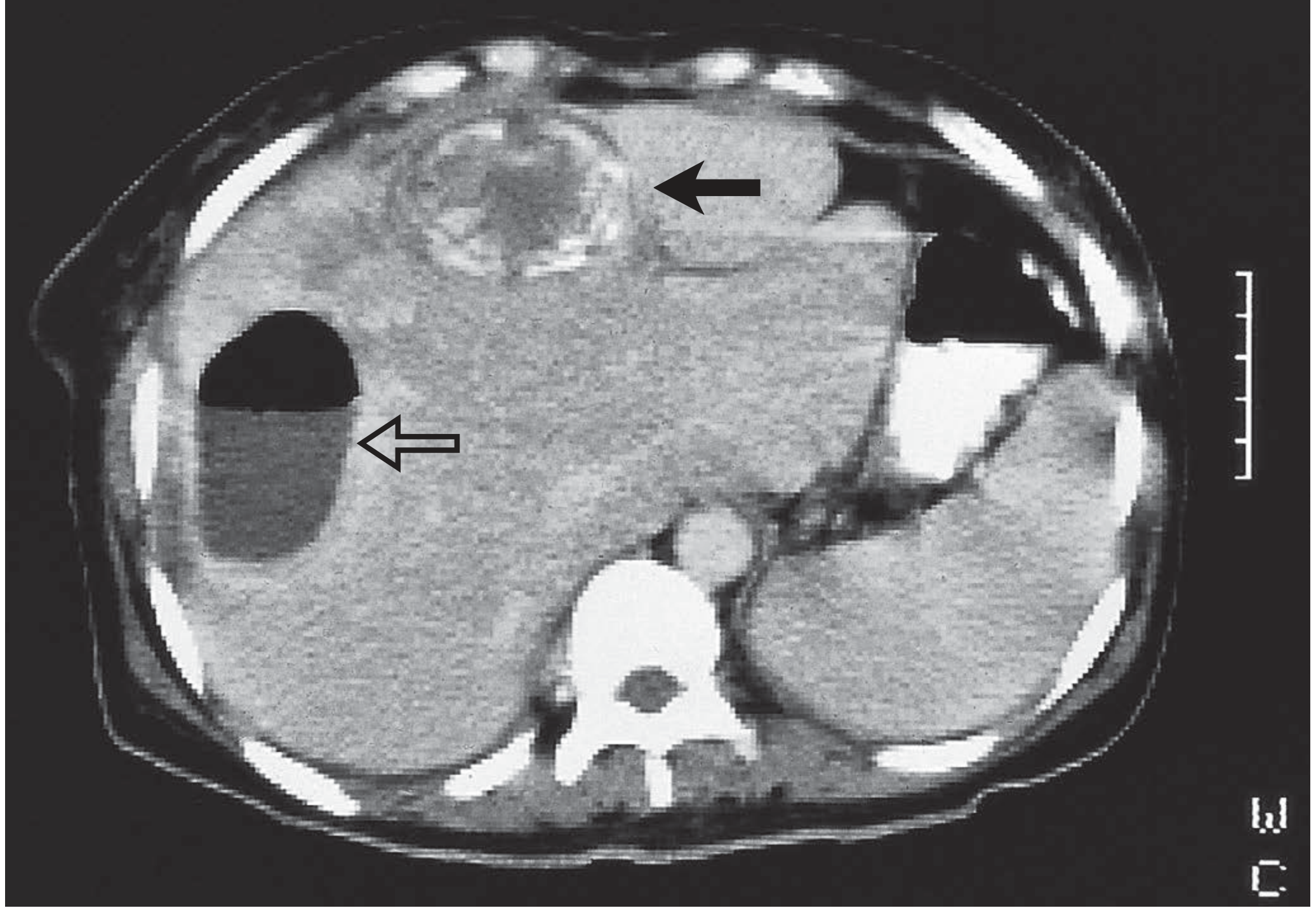
- CT abdomen: confirms diagnosis; shows rim enhancement + air-fluid level; sensitivity 95%+
- Aspiration: Gram stain, MC&S, Ziehl-Neelsen for TB, fungal culture
- Amoebic serology (ELISA >95% sensitive for amoebic abscess)
Management
Principles:
- Identify and treat the source
- Aspirate for microbiology (may require repeated aspiration)
- Antimicrobial therapy
Pyogenic liver abscess:
- IV antibiotics (combination):
- Metronidazole + clindamycin (anaerobic coverage, excellent cavity penetration)
- Third-generation cephalosporin or aminoglycoside (Gram-negative cover)
- Total duration: 4-6 weeks
- Percutaneous needle aspiration or catheter drainage (preferred for large/multiloculated)
- Treat underlying cause (e.g., ERCP for biliary source)
Amoebic liver abscess:
- Metronidazole 400-800 mg TDS x 10 days (drug of choice)
- Diloxanide furoate 500 mg TDS x 10 days (to eradicate intestinal carriage)
- Aspiration only if: no response to metronidazole after 72 hours, large abscess at risk of rupture, left lobe abscess (risk of peritoneal rupture)
Recurrent/refractory abscesses:
- Re-aspirate or insert drain
- Repeat microbiology
- Surgery (laparoscopy or open) if drainage and antibiotics fail
- Bailey and Love's, p. 2702-2720
QUICK REVISION SUMMARY TABLE
| Topic | Key Pathogen/Cause | Hallmark Finding | First-Line Tx |
|---|---|---|---|
| Acute diarrhoea | Rotavirus (children), Norovirus | Watery stools, dehydration | ORT ± antibiotics |
| Chronic liver disease | Alcohol, NAFLD, HBV | Spider naevi, ascites, varices | Treat cause, beta-blockers |
| Acute hepatitis | HAV, HBV, drugs | ALT 8-50x URL | Supportive; DAAs for HCV |
| NAFLD/MAFLD | Insulin resistance | AST:ALT <1, steatosis on USS | Weight loss, lifestyle |
| IBS | Gut-brain dysregulation | Rome IV criteria; no organic disease | Low FODMAP, antispasmodics |
| Gallstone disease | Cholesterol supersaturation | RUQ pain, Murphy's sign | Cholecystectomy |
| Liver abscess | Klebsiella, E. coli (pyogenic); E. histolytica (amoebic) | Fever, RUQ pain, multiloculated cyst | Antibiotics + aspiration |
Sources consulted:
- Goldman-Cecil Medicine International Edition (Elsevier)
- Bailey and Love's Short Practice of Surgery, 28th Edition
- Tietz Textbook of Laboratory Medicine, 7th Edition
- Robbins & Kumar Basic Pathology
- Sleisenger and Fordtran's Gastrointestinal and Liver Disease
- Yamada's Textbook of Gastroenterology, 7th Edition
- Harrison's Principles of Internal Medicine, 22nd Edition (2025)
- Medical Microbiology, 9th EditionAll 7 topics are now covered with detailed, exam-ready clinical notes. Here is a summary of what is included in each section:
What's Covered
1. Diarrhoea Diseases
- Mechanistic classification (secretory vs osmotic vs inflammatory vs motility)
- Acute vs chronic causes, red flags, investigations, and management including ORT principles and specific antibiotic therapy
2. Chronic Liver Disease / Cirrhosis
- Full pathobiology of hepatic stellate cell activation and portal hypertension (the NO paradox)
- All complications (varices, ascites, SBP, HRS, hepatic encephalopathy, HCC) with management
- Child-Pugh and MELD scoring, transplant criteria
3. Acute Hepatitis
- Direct vs immunologically-mediated injury distinction
- Full serology table for HAV/HBV/HCV
- Laboratory features table with AST:ALT ratios for each type
- King's College Criteria for liver transplant referral
4. Fatty Liver Disease
- NAFLD/MAFLD/NASH spectrum with complete pathogenesis (insulin resistance, two-hit model)
- Histological grading (NAS score)
- Key differentiator: AST:ALT <1 in NAFLD vs >2 in alcoholic liver disease
- Updated 2024 therapy options including resmetirom
5. Irritable Bowel Syndrome
- Full Rome IV diagnostic criteria with all subtypes
- Gut-brain interaction model, visceral hypersensitivity, post-infectious IBS
- Low FODMAP diet, stepwise pharmacotherapy by subtype
6. Gallstone Disease
- Stone types and risk factors, "5 Fs"
- Full clinical spectrum from biliary colic → acute cholecystitis → Charcot's triad → Rigler's triad
- Tokyo Guidelines diagnostic criteria for cholecystitis, management algorithms
7. Liver Abscess
- Pyogenic vs amoebic comparison table (including "anchovy sauce" aspirate for amoebic)
- CT image included
- Management: antibiotic combinations, drainage indications, amoebic treatment protocol
This is a shared conversation. Sign in to Orris to start your own chat.