
Intraamniotic haemorrhage
"intraamniotic hemorrhage" OR "intra-amniotic hemorrhage"
PMID: 27760979
PMID: 40898075
PMID: 29911274
PMID: 39631523
PMID: 17361084
intraamniotic haemorrhage causes diagnosis management obstetrics
https://discovery.ucl.ac.uk/10064801/1/Intraamniotic%20final…
intraamniotic hemorrhage ultrasound amniotic fluid echogenic
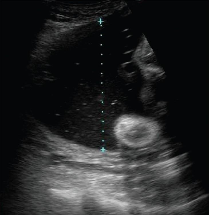
This obstetric diagnostic ultrasound image demonstrates the measurement technique for the single deepest vertical pocket (SDVP) of amniotic fluid. The primary feature is a large, predominantly anechoic (dark) area of liquor, which contains fine, low-level echogenic floating particles. These particles may represent vernix caseosa, though the differential diagnosis includes intraamniotic hemorrhage or infection (sludge). A vertical dotted line with 'plus' calipers illustrates the semiquantitative measurement of the fluid pocket, taken at a right angle to the uterine wall. In this clinical scenario, the large volume of fluid is consistent with hydramnios (polyhydramnios). Adjacent to the fluid pocket, a heterogeneous echogenic structure with a hyperechoic ring and hypoechoic center is visible, representing fetal or placental parts. The surrounding uterine and maternal tissues show varying degrees of echogenicity. This image is an educational example used in maternal-fetal medicine to teach the assessment of amniotic fluid volume and the identification of echogenic amniotic fluid.
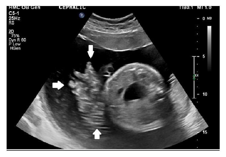
This obstetric ultrasound image, captured in a transverse plane, illustrates a rare clinical finding of active fetal hemorrhage. The central anatomical landmark is a cross-section of the fetal abdomen, characterized by a circular structure with a prominent anechoic (dark) internal region representing the fetal stomach or bladder. White arrows indicate a region of heterogeneous, echogenic swirling fluid within the normally anechoic amniotic cavity. This visual phenomenon represents blood emanating from the umbilical cord, which is seen adjacent to the fetal body. The echogenicity of the blood is notably higher than the surrounding amniotic fluid, indicating particulate matter (red blood cells) in motion. Superior to the fetus, the placental plate is visible as a moderately echogenic, curved boundary. This image is of high clinical significance for identifying spontaneous umbilical cord hemorrhage, a life-threatening obstetric emergency often associated with umbilical cord abnormalities such as a lack of Wharton's jelly or vessel exposure.
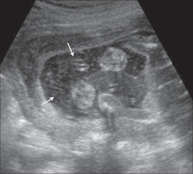
**Imaging Modality:** Obstetric Ultrasound (Gray-scale) **Anatomical Region:** Intrauterine environment, specifically the amniotic cavity. **Observed Pathology/Features:** The image demonstrates numerous fine, punctate, and linear hyperechoic particles (particulate matter) suspended within the normally anechoic (black) amniotic fluid. These floating internal echoes (indicated by white arrows) represent debris within the gestational sac. Surrounding fetal parts are visible but lack sharp definition due to the presence of the suspended material. **Characteristic Visual Features:** The distribution of the echoes is diffuse and heterogeneous, creating a "snowstorm" or turbid appearance within the fluid. There is no evidence of significant shadowing, suggesting a soft-tissue or cellular composition rather than calcification. **Clinical Context & Differentiation:** In this clinical presentation, the echogenic amniotic fluid is associated with fetal skin desquamation. Differential considerations for echogenic amniotic fluid typically include vernix caseosa (especially in the third trimester), intra-amniotic hemorrhage, or meconium staining. The specific postnatal diagnosis in this case is aplasia cutis congenita, where the echoes reflect sloughed cutaneous material.
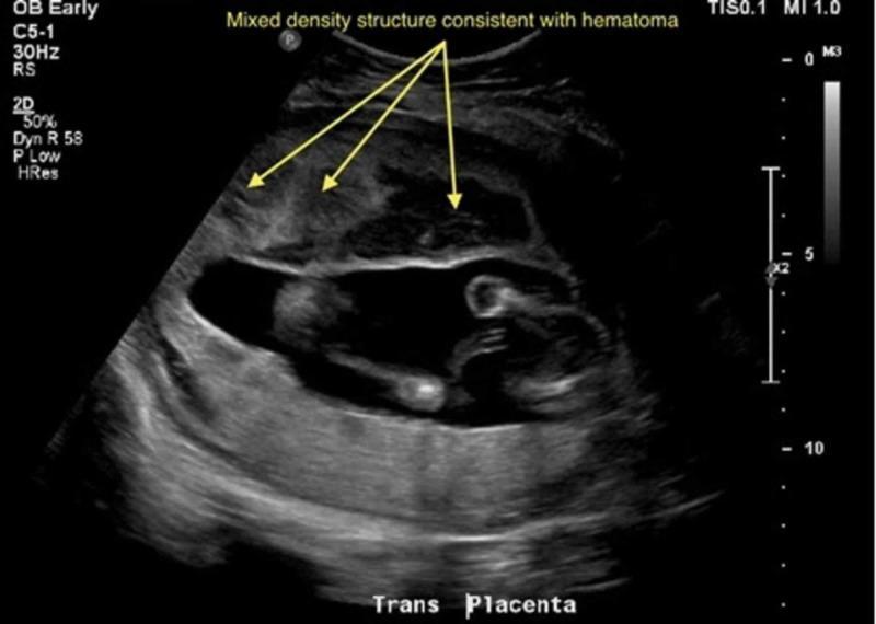
This obstetric diagnostic ultrasound image, oriented in a transverse plane ('Trans Placenta'), illustrates a significant pathology following trauma. The primary finding is a large, anteriorly located heterogeneous mass measuring approximately 12.0 x 5.4 x 9.5 cm. This mixed-density structure exhibits irregular echotexture, characterized by interspersed hypoechoic (darker) and echogenic (brighter) areas, indicated by yellow arrows and labeled as consistent with a hematoma. Below this collection, the amniotic cavity is visible, containing fetal structures and anecdotal evidence of amniotic fluid. In the lower (posterior) segment of the uterus, the placenta is visualized. The visual characteristics of the anterior heterogeneous mass, particularly its location relative to the placental site and its internal echoes, are highly suggestive of placental abruption with a retroplacental or preplacental hematoma. This image serves as a clinical teaching tool for evaluating obstetric trauma and identifying signs of acute placental separation and internal uterine hemorrhage in an emergency setting.
Intraamniotic Haemorrhage
Definition and Classification
- Maternal blood - from placental/uterine vessel disruption entering the amniotic space
- Fetal blood - from fetal vessels (cord, fetal surface vessels), representing a feto-amniotic haemorrhage
- Mixed - both sources simultaneously
Aetiology and Causes
| Category | Specific Causes |
|---|---|
| Placental | Placental abruption (concealed), circumvallate placenta (transplacental tear), placenta previa with accreta spectrum, placental laceration |
| Cord/Fetal vessels | Velamentous cord insertion, vasa praevia rupture, umbilical cord haemangioma, cord without Wharton's jelly |
| Iatrogenic | Amniocentesis, chorionic villus sampling, external cephalic version, intrauterine transfusion |
| Traumatic | Abdominal trauma |
| Spontaneous/Idiopathic | No identifiable cause found (rare - described at all gestations) |
Pathophysiology
- Fetal anaemia - if the source is fetal blood (can be confirmed by elevated MCA-PSV on Doppler)
- Polyhydramnios - blood draws fluid osmotically into the cavity; additionally, blood clots can mechanically obstruct flow
- PPROM - haematoma creates membrane tension and inflammatory mediators that degrade membranes; haemosiderin-laden macrophages are found in the chorion on histology
- Fetal distress - hypoxia from anaemia or cord compression by haematoma
- Maternal haemorrhagic shock - if blood loss is large enough (maternal source)
Clinical Presentation
- Antepartum vaginal bleeding (may be absent in fully concealed cases)
- Abdominal pain / uterine contractions
- Maternal cardiovascular compromise (tachycardia, hypotension, collapse)
- Decreased fetal movements
- Fetal heart rate abnormalities (reduced beat-to-beat variability, Category II/III tracing)
- Asymptomatic - discovered incidentally on routine ultrasound
Diagnosis
Ultrasound (First-line)
- Diffuse echogenic amniotic fluid ("snowstorm" appearance)
- Organised blood clot - heterogeneous echogenic mass, no Doppler flow within it (distinguishes from tumour/gastroschisis)
- Polyhydramnios - often accompanies the bleed
- MCA-PSV elevation - suggests fetal anaemia from fetal blood loss
- A freely mobile flap of placental tissue may be seen with circumvallate placenta tear
- Colour Doppler: absence of flow within the echogenic mass helps confirm haematoma
MRI (Problem-solving)
- Acute/subacute haemorrhage: T1 isointense, T2 hypointense amniotic fluid (paramagnetic effect of deoxyhaemoglobin/methaemoglobin)
- As blood ages, signal characteristics evolve predictably
- MRI reliably excludes gastroschisis and defines extent of haematoma
- First MRI description of concealed IAH in placenta previa with accreta spectrum was reported by Jha et al., Emerg Radiol 2018
Adjunct tests
- Kleihauer-Betke (KB) test or flow cytometry on maternal blood - quantifies fetal red cells in maternal circulation (feto-maternal haemorrhage)
- Abt's test (Apt test) on amniotic fluid - detects foetal haemoglobin (HbF) vs maternal HbA
- Elevated maternal serum AFP - reported in cases of IAH even without visible defect
- MCA-PSV Doppler - threshold >1.5 MoM for fetal anaemia
- FHR/CTG - assess for fetal compromise
- Full blood count - maternal anaemia in large maternal source bleeds
Differential Diagnosis
| Condition | Distinguishing features |
|---|---|
| Gastroschisis | Loops of bowel outside abdomen; Doppler may show flow; MRI confirms |
| Meconium-stained liquor | Clinical context (post-dates, fetal distress); echogenicity less marked |
| Vernix caseosa (third trimester) | Fine floating echoes, normal presentation near term |
| Intra-amniotic infection | Sludge/debris; clinical fever, WBC elevation; culture positive |
| Retroplacental haematoma | Located behind the placenta, not free in cavity |
| Subchorionic haematoma | Located at periphery under chorion |
| Vasa praevia rupture | Presents at membrane rupture with fetal bradycardia; APT test positive on vaginal blood |
Management
Conservative (selected cases at viable but preterm gestation)
- Hospital admission for monitoring
- Serial ultrasound and MCA-PSV Doppler
- Continuous CTG monitoring
- Betamethasone for fetal lung maturity if <34 weeks
- Treat underlying cause where identifiable
- A first-trimester case progressed to live birth at 31 weeks after PPROM and conservative management; histology showed haemosiderin-laden macrophages in chorion confirming the diagnosis
Active intervention
- Intrauterine transfusion - if fetal anaemia is confirmed (MCA-PSV >1.5 MoM) and gestation is preterm
- Delivery (emergency caesarean section) - indicated when:
- Maternal haemorrhagic shock
- Non-reassuring fetal heart rate not responding to conservative measures
- Gestation is near/at term
- Ongoing significant haemorrhage
Outcomes and Prognosis
- Fetal mortality is a real risk, particularly with fetal source bleeding, large haematoma compressing the cord, or severe anaemia
- Cases in the literature report live births with normal neonatal outcomes following timely intervention
- PPROM is a known complication from membrane damage by the haematoma
- Maternal morbidity depends on the volume of blood loss; large maternal bleeds can cause haemorrhagic shock
- Spontaneous resolution without adverse outcome has been reported in first-trimester cases with small haematomas
Summary
- Suspect it in any pregnancy with echogenic amniotic fluid on ultrasound, unexplained antepartum haemorrhage, or maternal collapse without an obvious cause
- Confirm with ultrasound (echogenic fluid, avascular mass, polyhydramnios) ± MRI
- Determine source (maternal vs fetal) with Apt/KB test and MCA-PSV Doppler
- Assess fetal wellbeing urgently with CTG and Doppler
- Manage based on gestational age and severity - conservative with close monitoring vs urgent delivery
Ultrasound Analysis - Intraamniotic Haemorrhage
Key Finding: Diffusely Echogenic Amniotic Fluid
Detailed Findings
| Feature | Finding | Significance |
|---|---|---|
| Amniotic fluid echogenicity | Diffusely echogenic with suspended particulate matter | Blood products in amniotic fluid |
| Organised clot | No large discrete avascular mass visible in this frame | May be early/diffuse bleed vs clot |
| Fetal parts | Hyperechoic fetal bone structures (limbs) visible | Fetus present |
| Placenta | Not clearly delineated in this scanning plane | Needs dedicated placental views |
| AFI/fluid volume | Fluid pocket appears present | Polyhydramnios needs AFI assessment |
Differential Diagnosis for Echogenic Amniotic Fluid
- Intraamniotic haemorrhage (most significant - given clinical context)
- Thick meconium staining (clinical context: post-dates, fetal distress)
- Severe intraamniotic infection/chorioamnionitis (fever, maternal tachycardia)
- Vernix caseosa (fine floating echoes, typically late third trimester, less dense)
What Additional Views Are Needed
- MCA-PSV Doppler - to detect fetal anaemia (threshold >1.5 MoM for gestational age)
- Colour Doppler of the amniotic cavity - absent flow within echogenic areas confirms haematoma vs vascular mass
- Placental assessment - look for abruption, circumvallate configuration, accreta spectrum
- Cord insertion site - rule out velamentous insertion or subamniotic haematoma
- CTG/fetal heart rate tracing - assess fetal wellbeing immediately
- Apt test / Kleihauer-Betke - determine if blood is fetal or maternal origin
Clinical Correlation Required
- Immediate CTG to assess fetal wellbeing
- MCA-PSV to quantify fetal anaemia
- Determine gestational age - to guide delivery vs conservative management
- Consider MRI if ultrasound is inconclusive about haematoma extent or differential diagnosis (e.g. gastroschisis)
- Urgent senior obstetric review
No fetal heart rate
Clinical Interpretation
- Diffusely echogenic amniotic fluid (intraamniotic haemorrhage confirmed on prior scan)
- Absent fetal heart rate
Likely Mechanism of Death
- Haemorrhage into the amniotic cavity (from fetal vessels, cord, or placenta)
- Fetal exsanguination - acute blood loss leading to profound anaemia and cardiovascular collapse
- OR cord compression by a blood clot causing acute hypoxia/asphyxia
- OR placental abruption with uteroplacental insufficiency
Immediate Management
Confirm the Diagnosis
- Real-time ultrasound - confirm absence of cardiac activity (must observe for at least 2 minutes in real time, not a single frozen frame)
- M-mode through the fetal heart - document asystole formally
- A second senior clinician should confirm
Inform the Family
- Sensitive, compassionate communication
- Avoid premature or uncertain statements - confirmation must be documented first
- Bereavement support team involvement
Maternal Assessment
- Full blood count - maternal anaemia if maternal source of bleed
- Coagulation screen - IUFD >4 weeks carries risk of DIC (consumptive coagulopathy from tissue thromboplastins); check fibrinogen, PT, APTT, D-dimer
- Kleihauer-Betke test - quantify feto-maternal haemorrhage (also relevant for anti-D prophylaxis if mother is Rh-negative)
- Blood group, crossmatch, renal function, LFTs
- Vital signs, haemodynamic status
Delivery Planning
- Vaginal delivery is preferred in most cases of IUFD (lower maternal morbidity than caesarean)
- Induction of labour with misoprostol or mifepristone + misoprostol depending on gestational age and local protocol
- If maternal haemorrhage/DIC is present, delivery is more urgent
- Caesarean only if specific obstetric indication (e.g. previous uterine surgery, maternal instability)
After Delivery - Investigations to Establish Cause
- Placenta - histopathology (circumvallate configuration, abruption, retroplacental clot, inflammatory changes)
- Cord - velamentous insertion, vessel anomaly, absent Wharton's jelly
- Fetal/neonatal post-mortem - with parental consent
- Genetics/karyotype if no clear cause found
- Apt test on amniotic fluid (if sample obtained) - confirms fetal vs maternal blood
DIC Risk in IUFD
| Time since death | DIC risk |
|---|---|
| < 4 weeks | Low |
| > 4 weeks | ~25-35% risk |
| With abruption | Elevated at any time |
Documentation
- Date and time of absent cardiac activity confirmed
- Names of two clinicians confirming
- Gestational age
- Last known fetal heart rate (if any)
- Ultrasound findings
History of trauma
Revised Diagnosis
Placental Abruption (most common traumatic cause)
- The placenta is inelastic - it cannot deform with the uterus during sudden deceleration/impact
- Shearing force at the uteroplacental interface → placental separation
- Blood tracks into the amniotic cavity (concealed abruption → intraamniotic haemorrhage)
- Even minor trauma can cause significant abruption - the severity of external injury does not correlate with degree of placental injury
- Abruption can occur up to 24-48 hours after trauma
Other Traumatic Mechanisms
| Mechanism | Notes |
|---|---|
| Direct uterine rupture | Rare; major blunt force; maternal signs of peritonism |
| Cord avulsion/laceration | Direct puncture, steering wheel injury |
| Feto-maternal haemorrhage | Placental tear → fetal blood into maternal circulation; can cause fetal exsanguination |
| Vascular injury to placental plate | Disruption of chorionic vessels |
Nature of Trauma - Critical to Establish
Accidental Trauma
- Road traffic accident (most common)
- Falls
- Workplace injury
- Direct abdominal blow (sports)
Non-Accidental Injury (Intimate Partner Violence / Domestic Abuse)
- Pregnancy is a high-risk period for domestic violence - incidence increases
- Abdominal targeting is characteristic of assault during pregnancy
- Must be screened for sensitively and privately
- IUFD from assault = potential criminal investigation
Safeguarding alert: If the history is inconsistent with the injury pattern, if there is delay in presentation, or if the partner is controlling the consultation, domestic abuse must be considered and local safeguarding protocols activated.
Additional Investigations in Trauma Context
Maternal
- CT abdomen/pelvis (or FAST ultrasound if unstable) - assess for splenic/hepatic/uterine injury, retroperitoneal haematoma
- Full trauma series - per ATLS protocol
- Coagulation screen + fibrinogen - abruption triggers DIC rapidly
- Kleihauer-Betke / flow cytometry - quantify feto-maternal haemorrhage; also determines anti-D dose if Rh-negative
- Urinalysis - haematuria suggests renal/bladder trauma
- Continuous maternal monitoring - at least 4-6 hours after any significant trauma in pregnancy (some guidelines say 24 hours if CTG abnormal)
For Cause of Death Documentation
- Placental histology - retroplacental clot, decidual necrosis, intervillous haemorrhage confirm traumatic abruption
- Post-mortem - confirms fetal exsanguination vs asphyxia vs direct fetal injury
- Photography of placenta at delivery - document clot, area of separation
- Weigh retroplacental clot if present
Medicolegal Considerations
| Issue | Action |
|---|---|
| Domestic violence suspected | Safeguarding referral; social work; police if consent given or mandatory reporting applies |
| Criminal injury (assault) | Preserve evidence; forensic documentation; chain of custody for samples |
| Road traffic accident | Police involvement as appropriate; injury documentation |
| Insurance/compensation claim | Detailed contemporaneous notes |
| Death certificate | Cause of death: IUFD secondary to intraamniotic haemorrhage due to placental abruption - traumatic aetiology |
Immediate Management Priorities
- Maternal stabilisation first - ATLS/ALSO protocol; airway, breathing, circulation
- Confirm IUFD as previously outlined
- Coagulation screen urgently - abruption + IUFD = high DIC risk; do not delay delivery if coagulopathy developing
- Establish nature and mechanism of trauma - history from patient alone (not in presence of partner/family)
- Activate safeguarding pathway if any concern about non-accidental injury
- Induction of labour - once maternal stabilisation achieved; avoid delay given DIC risk
- Multidisciplinary involvement - obstetrics, trauma surgery, haematology, social work, bereavement team