
Pain and stiffness in the right shoulder for the past 5 months, with difficulty performing overhead and behind-the-back activities.
adhesive capsulitis frozen shoulder anatomy diagram
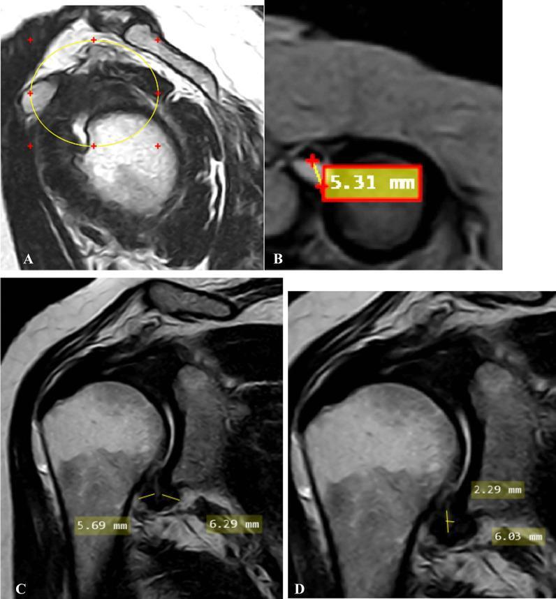
A multi-panel MRI study of the shoulder demonstrating radiological signs of adhesive capsulitis (frozen shoulder). Image A (oblique sagittal T2-weighted) shows the complete obliteration of the subcoracoid fat triangle within the rotator interval (encircled). Image B (oblique axial fat-suppressed PD-weighted) identifies a significant effusion (5.31 mm) within the long head of the biceps tendon sheath, exceeding the normal threshold. Images C and D (oblique coronal T2-weighted) focus on the axillary recess. Image C measures the pathological thickening of the joint capsule at the humeral (5.69 mm) and glenoid (6.29 mm) aspects. Image D provides measurements for the maximal height (2.29 mm) and width (6.03 mm) of the axillary recess space. This composite image illustrates key diagnostic criteria including rotator interval fat replacement, synovial effusion, and capsular thickening, which are critical for the radiological staging of adhesive capsulitis in a clinical setting.
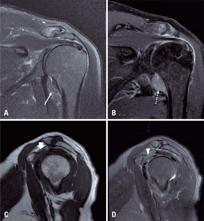
This diagnostic image set consists of four MRI views of a human shoulder illustrating the classic findings of adhesive capsulitis (frozen shoulder). Image A (coronal T2-weighted with fat suppression) and Image B (coronal T1-weighted post-gadolinium with fat suppression) demonstrate significant thickening of the glenohumeral joint capsule and pericapsular edema, particularly within the axillary recess. The post-contrast image (B) shows intense enhancement of the thickened capsule, indicating active synovitis and hypervascularity. Images C (sagittal T1-weighted) and D (sagittal T2-weighted with fat suppression) focus on the rotator interval. They reveal the characteristic obliteration of the normal subcoracoid fat pad by hypointense inflammatory tissue on T1 (arrow in C) and hyperintense edema on T2 (arrowhead in D). These imaging features are essential for confirming adhesive capsulitis in patients presenting with clinical shoulder stiffness and pain, allowing for the differentiation from other pathology such as rotator cuff tears or labral injuries.
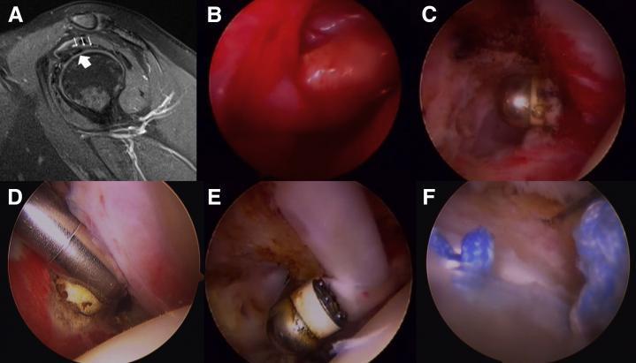
This composite image illustrates the diagnostic and surgical management of adhesive capsulitis (frozen shoulder). Panel A is a sagittal proton density fat-saturated MRI of the shoulder, demonstrating a thickened coracohumeral ligament (CHL, indicated by thin arrows) positioned superiorly to the long head of the biceps tendon (LHBT, indicated by a thick arrow), a characteristic radiological sign of this condition. Panels B through F provide a sequential arthroscopic view of the surgical intervention. Panel B shows severe synovitis with erythematous and thickened fibrous tissue obscuring the rotator interval. Panels C and D visualize the arthroscopic release of the rotator interval and the inferior glenohumeral ligament using specialized instruments. Panel E depicts the release of intra-articular adhesions surrounding the biceps tendon. Panel F shows the final procedural stage: a biceps tenodesis performed using a suture anchor technique, with visible blue sutures and the anchor site. The sequence highlights the progression from diagnostic imaging of ligamentous pathology to the intraoperative debridement and reconstructive stabilization of shoulder structures.
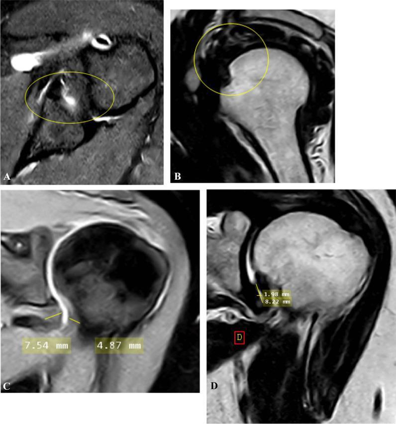
This composite of magnetic resonance imaging (MRI) scans illustrates diagnostic findings for adhesive capsulitis (frozen shoulder) across four planes. Panel A shows an oblique axial fat-suppressed proton density (PD)-weighted image demonstrating anterior and posterior extracapsular edema, indicated by hyperintense signals within the yellow circle. Panel B displays an oblique sagittal T2-weighted image showing partial obliteration of the subcoracoid fat triangle (encircled), a key indicator of proliferative soft tissue in the rotator interval. Panels C and D provide oblique coronal views used for quantitative assessment. Panel C (fat-suppressed PD) illustrates the measurement of the joint capsule's thickness at both the humeral and glenoid portions of the axillary recess (4.87 mm and 7.54 mm). Panel D (T2-weighted) shows the measurement of the axillary recess's maximal height and width (1.98 mm and 8.22 mm). These imaging modalities are critical for evaluating capsular thickening, edema, and recess volume loss in patients with clinical shoulder stiffness.
adhesive capsulitis frozen shoulder
Adhesive Capsulitis (Frozen Shoulder)
Clinical Diagnosis
- Global loss of active AND passive glenohumeral movement (not just active)
- Pathognomonic sign: loss of active external rotation
- Restricted shoulder motion such that the scapula moves en bloc with the arm during attempted movement
- Normal plain radiographs (distinguishes it from osteoarthritis)
- Pain often spontaneous in onset; minor trauma may be reported but its relevance is unclear
Pathophysiology
- Capsular thickening at the axillary recess
- Obliteration of the subcoracoid fat triangle (rotator interval changes)
- Synovial enhancement on contrast imaging
Associated Conditions / Risk Factors
- Females in their 50s (most common demographic)
- Diabetes mellitus (course is often prolonged and more severe)
- Thyroid disease
- Cardiac disease
- Immobility or neglect of a prior shoulder condition
- Depression and anxiety (adversely affect symptoms and function)
- Can complicate surgery or other painful shoulder conditions
Clinical Phases
| Phase | Features |
|---|---|
| Painful (Freezing) | Severe spontaneous pain, onset of stiffness, typically months 1-6 |
| Stiffening (Frozen) | Pain begins to ease, but stiffness peaks; ROM severely limited |
| Thawing | Progressive return of motion, pain continues to decrease |
Investigations
| Test | Finding |
|---|---|
| Plain X-ray shoulder | Normal (rules out OA, fracture) |
| MRI | Capsular thickening, axillary recess volume loss, rotator interval obliteration |
| Arthrography | Decreased capsular volume (not always needed) |
| Blood glucose / HbA1c | Screen for diabetes |
| Thyroid function tests | Screen for hypothyroidism |
Treatment
Conservative (First-line)
- Analgesia / NSAIDs - for pain control in the freezing phase
- Corticosteroid injections - into the glenohumeral joint and/or subacromial space; methylprednisolone acetate 40 mg/mL is a standard choice
- More commonly indicated in the stiffening phase
- Physiotherapy - begin with pendulum exercises and finger wall-climbing, progress to active ROM and strengthening
- Ice packs, ultrasound, TENS can supplement
- Distension injection (Hydrodilatation) - glenohumeral injection of corticosteroid combined with a large volume (20-30 mL) of local anaesthetic to distend the capsule; this facilitates subsequent physiotherapy
Operative (If Prolonged / Refractory)
- Manipulation Under Anaesthesia (MUA) with supplementary steroid injection
- Arthroscopic capsular release - release of the tight capsule (rotator interval, inferior glenohumeral ligament); useful for prolonged stiffness
- Note: A randomized trial found MUA + steroid injection, arthroscopic release, and structured physiotherapy were equivalently effective, but arthroscopic release carried the highest procedural risk - Goldman-Cecil Medicine
Summary Box (Bailey & Love)
- Most common in females in their fifties
- Spontaneous onset; severe pain with reduced glenohumeral motion
- Spontaneous resolution over 1-2 years
- Differentials: calcific tendinitis and rotator cuff tear
- Injections, distension with saline, manipulation, and surgical release may all help
Recent Evidence
Key Clinical Summary
- Confirm the diagnosis (clinical exam for global ROM loss, loss of external rotation; X-ray to exclude OA)
- Screen for diabetes and thyroid disease
- Initiate NSAIDs + glenohumeral corticosteroid injection
- Start physiotherapy with pendulum and wall-climbing exercises
- Counsel the patient that recovery typically takes 1-2 years; diabetes may prolong this
- Consider hydrodilatation if response to initial injection is suboptimal
- Reserve MUA or arthroscopic release for prolonged, refractory cases
Physiotherapy intervention in this case
physiotherapy exercise frozen shoulder adhesive capsulitis
PMID: 36861780
PMID: 42307090
frozen shoulder physiotherapy exercises pendulum wall climbing stretching

A composite of four clinical photographs demonstrating conventional physical therapy exercises for shoulder rehabilitation and range of motion (ROM) improvement. Top left: A patient performs 'wall washing' exercises, moving their hand in functional patterns against a wall surface to facilitate shoulder flexion and abduction. Top right: A patient uses a 'finger ladder' mounted on a wall, utilizing finger movements to incrementally increase shoulder elevation and abduction. Bottom left: A patient engages in shoulder pulley exercises using a suspended rope and handle system to perform assisted ROM activities, where the healthy arm helps lift the affected limb. Bottom right: A patient demonstrates a wall-climbing or stretching exercise to maximize vertical reach. These interventions are typically used in the management of adhesive capsulitis (frozen shoulder) or post-operative recovery to improve shoulder mobility, specifically targeting flexion, abduction, and rotation. The images represent standard physiotherapy protocols often compared with modern interventions like virtual reality-based therapy.
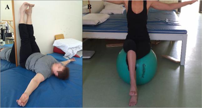
Clinical photographs depicting two rehabilitative exercises for patients with Charcot-Marie-Tooth (CMT) disease, aimed at managing symptoms through physiotherapy. Panel A illustrates a posterior chain muscle stretching technique. The patient is shown in a supine position on a treatment mat with both lower extremities elevated vertically and resting against a wall at a 90-degree angle. The arms are extended laterally to promote chest opening and stability, a position designed to address shortening of the posterior muscle chain. Panel B demonstrates a core stability exercise using a large green gymnastic ball (physioball). The patient is seated on the ball with arms abducted for balance while performing a single-leg lift, which engages the core musculature and improves proprioception and postural control. These images are representative of individualized physical therapy protocols designed to maintain mobility and enhance functional stability in neuromuscular disorders.
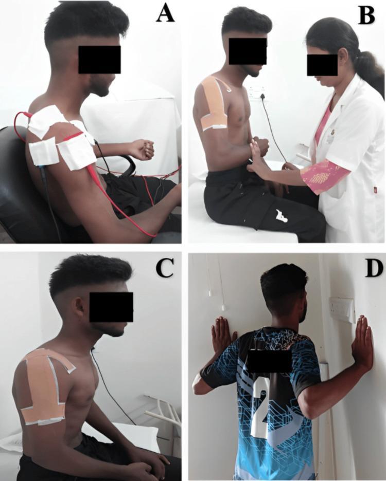
A multi-panel clinical photograph illustrating various stages of physiotherapy and rehabilitation for a shoulder injury. Panel A shows a seated patient receiving interferential therapy (IFT), with a four-pole electrode configuration using large white pads and red/black wires over the right shoulder and periscapular region for pain modulation. Panel B depicts a healthcare professional assisting the patient with isometric external rotation exercises; the patient's shoulder is stabilized with kinesiotherapy tape. Panel C focuses on specialized therapeutic taping, demonstrating an 'inverted J' application over the lateral and anterior deltoid aimed at increasing subacromial space. Panel D shows the patient performing active rehabilitation through wall push-ups in the scaption plane to improve scapular stability and muscle strength. The sequence represents a comprehensive orthopedic rehabilitation protocol ranging from passive pain management to active functional strengthening. The patient is a young adult male, and the setting is a clinical physiotherapy environment.
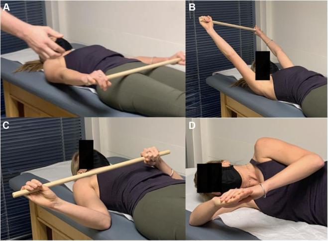
This composite of clinical photographs demonstrates various scapular and shoulder stretching exercises used in the rehabilitation of Pectoralis Minor Syndrome (PMS) and neurogenic thoracic outlet syndrome (NTOS). (A, B) Shoulder flexion stretch: The patient is in a supine position, holding a wooden stick with both hands. This allows the asymptomatic arm to assist the affected arm into maximal overhead flexion, targeting the anterior shoulder and pectoralis minor muscle. (C) External rotation stretch: Also performed supine using a stick, this exercise uses the contralateral arm to facilitate maximal external rotation, stretching the anterior shoulder anatomy. (D) Sleeper stretch: The patient is in a lateral decubitus position (lying on the affected side) with the arm flexed at the elbow. The patient uses the opposite hand to gently push the forearm toward the table, performing passive internal rotation to stretch the posterior shoulder capsule and associated anatomy. These exercises are critical for improving scapular kinematics and relieving neurovascular compression in the retropectoralis minor space.
PMID: 37981583
Physiotherapy Intervention for Adhesive Capsulitis (Frozen Shoulder)
Phase-Based Approach
Phase 1 - Painful (Freezing) Phase: Pain Control First
Electrophysical Modalities (Passive)
| Modality | Purpose | Notes |
|---|---|---|
| TENS (Transcutaneous Electrical Nerve Stimulation) | Analgesia via gate control | High-frequency TENS for acute/subacute pain |
| Therapeutic Ultrasound | Deep heating, tissue extensibility | Pulsed mode initially; continuous as pain settles |
| High-Intensity Laser Therapy (HILT) | Analgesia, disability reduction | A 2023 meta-analysis (PMID 37981583) showed HILT significantly reduces pain (VAS MD -2.23 cm) and disability (SPADI MD -10.1%) vs. conventional therapy alone, though it does not outperform conventional PT for ROM gains |
| Interferential Therapy (IFT) | Deep analgesia, muscle stimulation | Applied over the shoulder/periscapular region |
| Superficial heat / cold packs | Pain relief, muscle relaxation | Ice preferred in acute inflammatory phase; heat as pain settles |
| Shortwave Diathermy (SWD) | Deep heating of capsule | Useful in stiffening phase to improve extensibility before stretching |
Phase 2 - Stiffening/Frozen Phase: Mobility Restoration
Exercise Progression
Step 1 - Pendulum (Codman) Exercises
- Patient leans forward, arm hangs freely
- Uses trunk/gravity to initiate small circular or pendular movements
- Goal: Gentle distraction of glenohumeral joint; low-load sustained stretch; reduces protective muscle guarding
- Performed multiple times daily (3-5 sets, 1-2 min each)
Step 2 - Passive / Active-Assisted ROM
- Finger wall-walking: Patient faces a wall, walks fingers up to progressively increase flexion/abduction
- Pulley exercises: Unaffected arm assists the affected arm through flexion arc
- Stick/wand exercises: Unaffected arm uses a stick/rod to assist ER, flexion, and overhead movements
- Supine passive flexion: Therapist-assisted or gravity-assisted ROM in supine (less painful than upright)
- Cross-body adduction stretch: Targets posterior capsule
Step 3 - Capsular Stretching (Key for Adhesive Capsulitis)
| Stretch | Capsular Region Targeted |
|---|---|
| Inferior capsule stretch (overhead reach in supine) | Inferior glenohumeral ligament / axillary pouch |
| Posterior capsule stretch / Sleeper stretch | Posterior capsule; addresses limited IR |
| Anterior capsule stretch (ER stretch at 90° abduction) | Anterior band of inferior glenohumeral ligament |
| Cross-body stretch | Posterior capsule |
Manual Therapy
| Technique | Description |
|---|---|
| Maitland Joint Mobilization (Grades I-II) | Oscillatory glides within pain-free range; used in painful phase for neurophysiological analgesia |
| Maitland Grades III-IV | Larger amplitude glides into resistance/pain; used in frozen phase for capsular stretch and ROM gain |
| Inferior glenohumeral glide | With arm in slight abduction - opens the inferior capsule; most effective for restoring abduction |
| Posterior glenohumeral glide | Restores flexion and IR |
| Anterior glenohumeral glide | Restores ER |
| Mulligan MWM (Mobilization with Movement) | Sustained accessory glide combined with active movement; can achieve meaningful ER gains in one session |
| Soft tissue therapy / myofascial release | Targets periscapular muscles (upper trapezius, levator scapulae, pectoralis minor) that become overactive |
| Scapulothoracic mobilization | Improves overall shoulder girdle mechanics when glenohumeral motion is blocked |
Phase 3 - Thawing Phase: Strength and Function Restoration
Strengthening Progression
- Isometric exercises (first - pain-free): ER, IR, abduction, flexion against wall/doorframe
- Isotonic exercises with resistance band / Theraband:
- ER at side and at 90° abduction
- IR
- Scaption (shoulder elevation in the scapular plane) - recruits supraspinatus and deltoid with less impingement
- Rows, scapular retraction
- Rotator cuff strengthening: Systematic progression targeting supraspinatus, infraspinatus, teres minor, subscapularis
- Scapular stabilizers: Serratus anterior (wall push-ups in scaption plane), lower/middle trapezius (prone Y/T/W exercises)
Adjuncts and Combination Approaches
- Post-injection physiotherapy: After corticosteroid or hydrodilatation injection, commence PT within 24-48 hours while the capsule is more pliable - this window is critical for ROM gains
- Heat before stretching: Apply SWD/ultrasound or moist heat to the shoulder 10-15 min before capsular stretching to increase tissue extensibility
- Kinesiotaping: Applied over the deltoid in an "inverted J" pattern to increase subacromial space during active rehab
- Psychological support / relaxation: A 2026 meta-analysis (PMID 42307090) confirmed anxiety is consistently associated with frozen shoulder severity and adversely affects pain, function, and quality of life - addressing psychosocial factors is part of a biopsychosocial approach
Home Exercise Program (HEP)
- Pendulum exercises: 3-5 times/day
- Capsular stretching: at least twice daily, after a warm shower
- Wand/pulley exercises: daily
- Progressive ROM diary to track improvement
Overall Evidence Summary
| Intervention | Evidence Level | Effect |
|---|---|---|
| Exercise (any type) | Low-very low | Small but significant improvements in pain, function, ROM (PMID 42307090) |
| Manual therapy + exercise | Low-very low | Non-significant pooled effects; heterogeneous designs (PMID 36861780) |
| HILT + PT vs PT alone | Moderate | Significant pain and disability reduction; no ROM superiority (PMID 37981583) |
| Corticosteroid injection + PT | Moderate | Better short-term outcomes than PT alone (supported by RCTs) |
| Biopsychosocial model | Very low | Metabolic and psychological factors associated with severity (PMID 42307090) |
Key Clinical Points
- Never push aggressively in the painful phase - this increases inflammation and patient guarding
- The inferior capsule (axillary pouch) is the primary site of contracture - prioritize inferior and posterior glides
- External rotation loss is the most characteristic deficit - restoring ER is the primary ROM goal
- Diabetes prolongs the course and reduces response to treatment - metabolic control should run parallel to PT
- Total PT course: typically 6-12 weeks of supervised sessions with ongoing HEP; may extend to 12 months in refractory cases
- If no meaningful progress after 3-6 months of structured physiotherapy, escalate to hydrodilatation, MUA, or arthroscopic release