Explain me antrochoanla polyp in a easier way

Reading File
Finding Sources
Searching Images

antrochoanal polyp nasal endoscopy CT scan
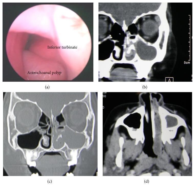
This composite clinical figure illustrates an antrochoanal polyp (ACP) through multi-modal imaging: endoscopy and computed tomography (CT). Panel (a) shows an endoscopic view of the left nasal cavity, identifying a smooth, pale mass located inferiorly at the level of the inferior meatus, adjacent to the inferior turbinate. Panel (b) is a coronal CT scan in a soft tissue window demonstrating a large cystic-appearing mass occupying the maxillary antrum. Panel (c) provides a coronal bone window CT, highlighting a focal defect in the medial wall of the maxillary sinus situated below the inferior turbinate, serving as the exit point for the polyp into the nasal cavity. Panel (d) displays an axial CT scan where the soft tissue mass is seen extending posteriorly from the nasal cavity into the nasopharynx. This sequence demonstrates the characteristic tri-compartmental progression of an ACP from the maxillary sinus, through the nasal meatus, and into the choana/nasopharynx, including rare inferior meatus involvement.
This composite clinical figure illustrates an antrochoanal polyp (ACP) through multi-modal imaging: endoscopy and computed tomography (CT). Panel (a) shows an endoscopic view of the left nasal cavity, identifying a smooth, pale mass located inferiorly at the level of the inferior meatus, adjacent to the inferior turbinate. Panel (b) is a coronal CT scan in a soft tissue window demonstrating a large cystic-appearing mass occupying the maxillary antrum. Panel (c) provides a coronal bone window CT, highlighting a focal defect in the medial wall of the maxillary sinus situated below the inferior turbinate, serving as the exit point for the polyp into the nasal cavity. Panel (d) displays an axial CT scan where the soft tissue mass is seen extending posteriorly from the nasal cavity into the nasopharynx. This sequence demonstrates the characteristic tri-compartmental progression of an ACP from the maxillary sinus, through the nasal meatus, and into the choana/nasopharynx, including rare inferior meatus involvement.

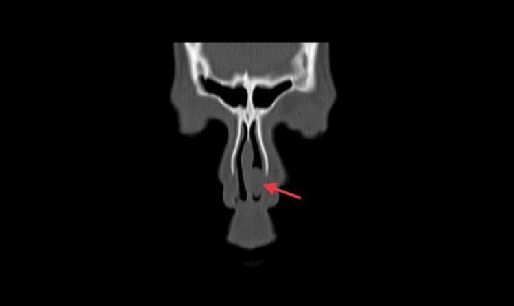
This diagnostic image is a non-enhanced coronal CT scan of the paranasal sinuses demonstrating a large antrochoanal polyp. The key visual finding is a significant soft tissue density (opacification) completely filling the left maxillary sinus and extending through a widened maxillary ostium into the left nasal cavity. A red arrow highlights a large, well-defined polypoidal mass within the left nasal fossa, which is causing local mass effect. Secondary findings include a significant deviation of the nasal septum toward the right side and compensatory hypertrophy of the right inferior turbinate (concha). The left inferior turbinate appears displaced and indented by the encroaching polypoidal mass. The sphenoid and frontal sinuses appear clear on this section. This image serves as a classic educational example of sinonasal polyposis and obstructive nasal pathology, illustrating the characteristic 'dumbbell' appearance of an antrochoanal polyp as it herniates from the sinus antrum into the nasal passage.
This diagnostic image is a non-enhanced coronal CT scan of the paranasal sinuses demonstrating a large antrochoanal polyp. The key visual finding is a significant soft tissue density (opacification) completely filling the left maxillary sinus and extending through a widened maxillary ostium into the left nasal cavity. A red arrow highlights a large, well-defined polypoidal mass within the left nasal fossa, which is causing local mass effect. Secondary findings include a significant deviation of the nasal septum toward the right side and compensatory hypertrophy of the right inferior turbinate (concha). The left inferior turbinate appears displaced and indented by the encroaching polypoidal mass. The sphenoid and frontal sinuses appear clear on this section. This image serves as a classic educational example of sinonasal polyposis and obstructive nasal pathology, illustrating the characteristic 'dumbbell' appearance of an antrochoanal polyp as it herniates from the sinus antrum into the nasal passage.

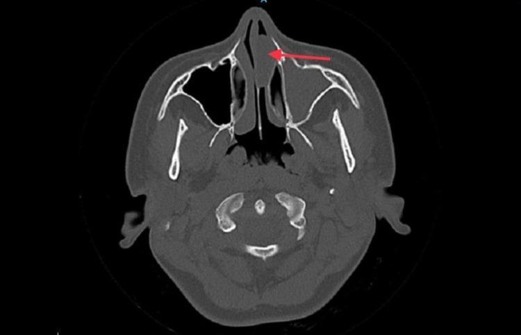
This diagnostic image is a non-contrast axial CT scan of the paranasal sinuses at the level of the maxillary antrum. The scan reveals a large, well-defined soft tissue density mass (indicated by a red arrow) within the left nasal cavity, characteristic of an antrochoanal polyp. This polypoidal lesion originates from the left maxillary sinus, which demonstrates complete opacification and widening of the maxillary ostium. There is evidence of mild bony remodeling of the left maxillary sinus walls. The mass extends anteriorly, causing significant obstruction of the left nasal passage and lateral displacement of the left inferior turbinate. Secondary findings include a marked diffuse deviation of the nasal septum toward the right side and compensatory hypertrophy of the right inferior turbinate. In contrast, the right maxillary sinus is well-aerated with normal mucosal thickness and clear ostiomeatal complex structures. This image serves as a clinical example of sinonasal polyposis and mechanical obstruction of the nasal airway, relevant for otolaryngology and radiology education.
This diagnostic image is a non-contrast axial CT scan of the paranasal sinuses at the level of the maxillary antrum. The scan reveals a large, well-defined soft tissue density mass (indicated by a red arrow) within the left nasal cavity, characteristic of an antrochoanal polyp. This polypoidal lesion originates from the left maxillary sinus, which demonstrates complete opacification and widening of the maxillary ostium. There is evidence of mild bony remodeling of the left maxillary sinus walls. The mass extends anteriorly, causing significant obstruction of the left nasal passage and lateral displacement of the left inferior turbinate. Secondary findings include a marked diffuse deviation of the nasal septum toward the right side and compensatory hypertrophy of the right inferior turbinate. In contrast, the right maxillary sinus is well-aerated with normal mucosal thickness and clear ostiomeatal complex structures. This image serves as a clinical example of sinonasal polyposis and mechanical obstruction of the nasal airway, relevant for otolaryngology and radiology education.

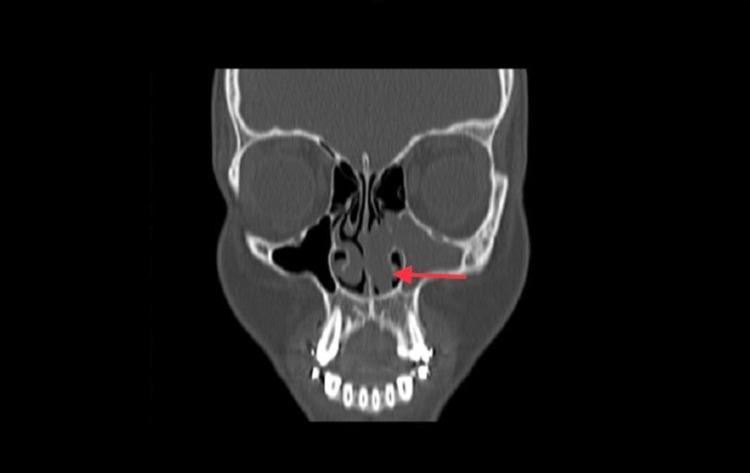
This diagnostic image is a non-contrast coronal CT scan of the paranasal sinuses demonstrating a large left-sided antrochoanal polyp. The left maxillary sinus shows total opacification and mild bony remodeling. This opacification extends through a significantly widened left maxillary ostium (indicated by a red arrow) into the left nasal cavity, representing the typical 'dumbbell' configuration of an antrochoanal polyp. Within the nasal cavity, a large polypoidal soft tissue mass obstructs the middle and inferior meatus, causing displacement of the left inferior turbinate and partial opacification of the middle meatus. The left uncinate process and hiatus semilunaris are obscured by mucosal thickening. The nasal septum is deviated toward the right. In contrast, the right maxillary sinus, ostium, and ethmoid air cells are clear and well-pneumatized, providing a healthy anatomical comparison. This image serves as a classic educational example of sinonasal polyposis and obstructive sinus pathology in otolaryngology.

This diagnostic image is a non-contrast coronal CT scan of the paranasal sinuses demonstrating a large left-sided antrochoanal polyp. The left maxillary sinus shows total opacification and mild bony remodeling. This opacification extends through a significantly widened left maxillary ostium (indicated by a red arrow) into the left nasal cavity, representing the typical 'dumbbell' configuration of an antrochoanal polyp. Within the nasal cavity, a large polypoidal soft tissue mass obstructs the middle and inferior meatus, causing displacement of the left inferior turbinate and partial opacification of the middle meatus. The left uncinate process and hiatus semilunaris are obscured by mucosal thickening. The nasal septum is deviated toward the right. In contrast, the right maxillary sinus, ostium, and ethmoid air cells are clear and well-pneumatized, providing a healthy anatomical comparison. This image serves as a classic educational example of sinonasal polyposis and obstructive sinus pathology in otolaryngology.

Reading File
Reading File
Loading Image
Here is a simple, easy-to-understand explanation of Antrochoanal Polyp (ACP):

Antrochoanal Polyp (ACP) - Made Simple

What Is It? (The Simple Analogy)

Think of your cheek (maxillary sinus) as a balloon inside a room. Normally it just sits there. In an antrochoanal polyp, that balloon grows so big it squeezes out through a small hole (the sinus opening), travels through the nose, and dangles all the way back into your throat.
That journey - from the antrum (maxillary sinus/cheek bone air space) to the choana (back opening of the nose into the throat) - is exactly where the name comes from:
  • Antro = antrum (maxillary sinus)
  • Choanal = choana (back of the nose/throat junction)

Where Does It Start?
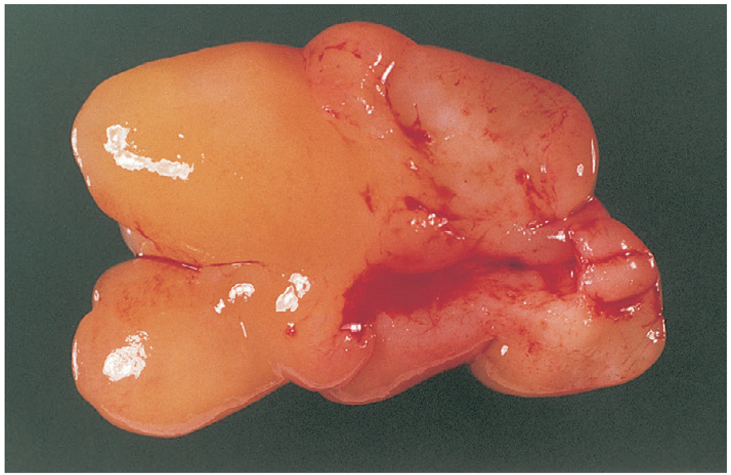
It starts as a benign (non-cancerous) fleshy growth from the lining of the maxillary sinus - the hollow air space inside your cheekbone. As it grows, it squeezes through the natural opening of the sinus (called the ostium or posterior fontanelle), enters the nasal passage, and eventually flops down into the nasopharynx (the area behind your nose where it connects to your throat).
This is the classic "dumbbell" shape - a part inside the sinus + a narrow neck through the opening + a bigger part hanging in the nose/throat.
Antrochoanal polyp - gross specimen after surgical removal
Antrochoanal polyp - gross specimen after removal (Bailey & Love's Surgery)

Key Features at a Glance

FeatureDetail
OriginMaxillary sinus (cheek sinus)
DirectionGrows backward toward the throat
Side affectedAlmost always ONE side only (unilateral)
NumberAlways a SINGLE polyp
Who gets itMore common in children & young adults (unlike regular nasal polyps which are in older adults)
Shape on CT"Dumbbell" - two bulges connected by a narrow neck

How Is It Different from Normal Nasal Polyps?

Regular Nasal PolypsAntrochoanal Polyp
OriginEthmoid sinuses (between eyes)Maxillary sinus (cheek)
NumberMultipleSingle
SideBoth sides (bilateral)One side only
Age groupMiddle-aged adultsChildren & young adults
Appearance on scopePale, grape-like clustersSingle large fleshy mass going back toward throat

Symptoms - What Does the Patient Feel?

Because the polyp grows into the nose and blocks it:
  • One-sided nasal blockage (can't breathe through one nostril)
  • Feeling of something at the back of the throat - the patient may notice a lump dangling in the throat
  • Runny nose, postnasal drip
  • In children: may cause snoring or mouth breathing
  • Pain is usually NOT a prominent feature

What Does It Look Like on Imaging?

Here's what a CT scan shows - the characteristic "dumbbell":
Multi-modal imaging of antrochoanal polyp showing endoscopy and CT scan views
Endoscopy and CT demonstrating a left-sided antrochoanal polyp with its tri-compartmental progression: maxillary sinus → nasal cavity → nasopharynx
Coronal CT showing antrochoanal polyp with widened maxillary ostium and dumbbell configuration
Coronal CT: the polyp fills the maxillary sinus and extends through a widened ostium into the nasal cavity - classic "dumbbell" appearance

Treatment - How Is It Treated?

Surgery is the treatment - medicines alone (steroids) don't cure it.
The key operation is Functional Endoscopic Sinus Surgery (FESS):
  1. A camera is inserted into the nose (no external cuts)
  2. The surgeon removes not just the nose part, but also the part inside the maxillary sinus - this is the most important step
  3. If the antral (sinus) portion is left behind, it WILL come back (recur)
The traditional open surgery (Caldwell-Luc - entering from under the upper lip) is no longer routinely needed, as per [Cummings Otolaryngology](modern FESS approaches).

Simple Summary in 3 Lines

An antrochoanal polyp is a single, benign fleshy lump that starts in the maxillary (cheek) sinus, squeezes through the sinus opening into the nose, and hangs backwards into the throat. It mainly affects one side, mostly in young people, and causes one-sided nasal blockage. Treatment is surgical removal (FESS), and to prevent recurrence, the part inside the sinus must be completely removed.

Sources: Bailey & Love's Short Practice of Surgery (28th ed.) | Cummings Otolaryngology Head & Neck Surgery
This is a shared conversation. Sign in to Orris to start your own chat.