
Strongyloides stercoralis
Reading File
Finding Sources
Searching Images
Strongyloides stercoralis larvae rhabditiform filariform
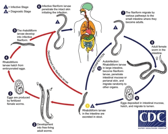
Summary : This diagram illustrates the life cycle of Strongyloides stercoralis, a parasitic nematode, showing both free-living and parasitic cycles, as well as the possibility of autoinfection in humans. The process includes stages in the environment and within the human host, with clear indications of infective and diagnostic stages.
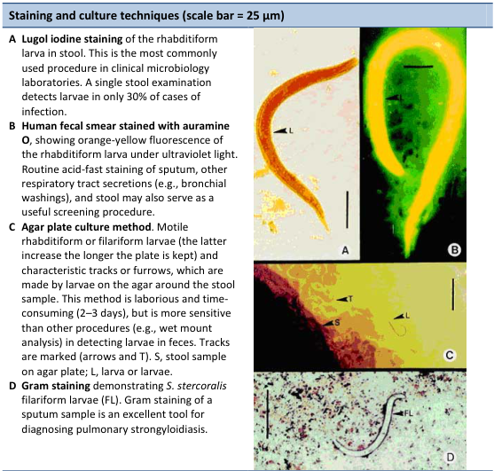
Summary : This figure presents four laboratory techniques for detecting Strongyloides stercoralis larvae in clinical samples, with corresponding micrographs illustrating each method. The scale bar in all images is 25 µm.
Searching PubMed
"Strongyloides stercoralis" AND treatment
Reading File
Reading File
Reading File
Reading File
Finding Sources
I have sufficient material now to give a thorough, well-organized answer.
Reading File
Strongyloides stercoralis
Overview
S. stercoralis ("human threadworm") is a soil-transmitted intestinal and tissue nematode notable for its unique ability to complete its entire life cycle within a single host through autoinfection, allowing infection to persist for decades without re-exposure.
Life Cycle
- Adult females (2-3 mm) live embedded in the duodenal/jejunal mucosa and reproduce parthenogenetically (no males needed in the parasitic phase).
- Eggs hatch in situ in the small bowel, releasing rhabditiform (L1) larvae, which pass in stool (eggs themselves are rarely seen on stool O&P).
- Direct cycle: L1 larvae molt in soil into infective filariform (L3) larvae, which penetrate intact skin (often bare feet), migrate via the bloodstream to the lungs, are coughed up and swallowed, then mature into adults in the small intestine.
- Indirect (free-living) cycle: under favorable soil conditions, larvae can develop into free-living, sexually reproducing adults, whose progeny again become infective filariform larvae.
- Autoinfection (the clinically critical feature): rhabditiform larvae can transform into filariform larvae within the intestine or on perianal skin and reinvade the host directly, without ever leaving the body - sustaining infection indefinitely.
Clinical Manifestations
- Cutaneous: pruritic, erythematous rash at larval entry site; chronic autoinfection can cause larva currens - rapidly migrating (up to 10 cm/hr), serpiginous urticarial tracks, typically on the trunk/buttocks/thighs.
- Pulmonary: larval migration through the lungs can cause cough, wheeze, and Löffler-type syndrome with peripheral eosinophilia.
- Gastrointestinal: epigastric pain resembling peptic ulcer disease, diarrhea, nausea, and, with heavy chronic infection, a malabsorption-like syndrome.
- Hyperinfection syndrome / disseminated strongyloidiasis: in immunosuppressed hosts (corticosteroid use, HTLV-1 infection, transplant recipients, chemotherapy, malnutrition, alcoholism), massively accelerated autoinfection produces overwhelming larval burden with severe GI hemorrhage/diarrhea, pulmonary hemorrhage, and dissemination to the CNS (larvae can carry enteric gram-negative bacteria into the bloodstream, causing secondary bacteremia/sepsis or meningitis). This is often fatal if unrecognized.
Diagnosis
- Stool microscopy: identification of rhabditiform larvae (short buccal cavity, prominent genital primordium, distinguishing them from hookworm larvae). Sensitivity of a single stool exam is low; up to 7 serial specimens may be needed, or agar plate/coproculture methods for higher sensitivity.
- Duodenal aspirate or string test if stool exams are unrevealing.
- Filariform larvae in sputum/bronchial wash/other extraintestinal fluid signals hyperinfection/dissemination - an urgent finding requiring immediate clinical notification.
- Serology (EIA): good sensitivity/specificity but cross-reacts with other nematode infections (e.g., filariasis) and cannot reliably distinguish past from current infection; useful for screening at-risk/immunosuppromised patients before immunosuppression and for monitoring treatment response.
- Peripheral eosinophilia is a common but nonspecific clue.
Treatment
- Ivermectin is the treatment of choice for uncomplicated strongyloidiasis (single or two-dose regimens); albendazole and thiabendazole are alternatives, though generally less effective than ivermectin.
- Hyperinfection/disseminated disease: prolonged ivermectin (often until larvae clear from stool/sputum and symptoms resolve, sometimes with repeated dosing), and in patients who cannot absorb oral therapy, subcutaneous (veterinary-formulation) ivermectin has been used as salvage therapy.
- Screening and empiric/pre-emptive treatment is recommended before starting immunosuppressive therapy (e.g., corticosteroids, transplant, chemotherapy) in patients from endemic areas or with unexplained eosinophilia, to prevent hyperinfection.
Sources
- Jawetz, Melnick & Adelberg's Medical Microbiology, 28th ed.
- Henry's Clinical Diagnosis and Management by Laboratory Methods
- Robbins, Cotran & Kumar Pathologic Basis of Disease
- Murray & Nadel's Textbook of Respiratory Medicine
Recent evidence check
A 2024 systematic review comparing ivermectin vs moxidectin for treating S. stercoralis infection (PMID: 39819639) and a 2019 multicenter RCT (Strong Treat 1-4, cited in Goldman-Cecil Medicine) support ivermectin's continued first-line role, with moxidectin emerging as a comparably effective alternative in ongoing trials - this doesn't contradict the textbook guidance above but reflects an evolving option worth watching. A 2024 UK systematic review/meta-analysis (PMID: 39009349) also reinforces that strongyloidiasis remains underdiagnosed outside classic endemic regions, consistent with the textbook note about non-tropical foci (e.g., historically in parts of Appalachia).
This is a shared conversation. Sign in to Orris to start your own chat.