
Radiology of meningioma from Grainger and osborne
Reading File
Meningiomas are extra-axial, dural-based tumors that account for ~15-20% of intracranial tumors. On imaging they show characteristic features: isodense on plain CT, homogeneous enhancement, calcification in ~25%, and a "dural tail." On MRI they are iso- to hypointense on T1, variable on T2, with avid enhancement.
Searching sources...
Finding Sources
Finding Sources
Found it - Grainger & Allison's diagnostic radiology (ISBN 9780323760751). Now let me read the meningioma section and search for more related content in that book.
Reading File
Finding Sources
There's a dedicated meningioma section in this book - let me search for the main extra-axial/extraventricular meningioma content which is referenced as "see below."
Finding Sources
Finding Sources
Found the main MENINGIOMAS section. Now let me read it fully along with the optic nerve sheath meningioma section and the spinal meningioma section.
Reading File
Reading File
Excellent content found. Now let me read more of the main MENINGIOMAS section which was truncated, and also get the spinal meningioma section and fetch the key images.
Reading File
Reading File
I have substantial content. Now let me read the continuation of the meningioma section (physiological MRI and advanced imaging) and fetch the key figures.
Reading File
Loading Image
Loading Image
I now have comprehensive content from Grainger & Allison's. Let me fetch the remaining key images (the PET and spinal images) to complete the visual set.
Loading Image
Loading Image
I now have all the key content and images from Grainger & Allison's. Let me compile the comprehensive answer.
Radiology of Meningioma
Source: Grainger & Allison's Diagnostic Radiology
Overview
Meningiomas originate from meningocytes or arachnoid cell rests related to arachnoid granulations of the dura mater. They represent the commonest non-glial intracranial neoplasm, accounting for approximately 20% of all primary intracranial tumours. They may be spherical and well-circumscribed, or flat and infiltrating ("en plaque" lesions).
WHO Classification
| Grade | Type | Features |
|---|---|---|
| I | Typical | Good prognosis, low recurrence after resection |
| II | Atypical | High mitotic index, disordered growth, or brain invasion |
| III | Anaplastic | Excessive mitotic activity; sarcoma/carcinoma/melanoma-like appearance |
Location
- 90% are supratentorial, arising in decreasing frequency from:
- Parasagittal region
- Cerebral convexities
- Sphenoid ridge
- Olfactory groove
- Infratentorial meningiomas are most frequent on the posterior surface of the petrous bones and clivus; can mimic vestibular schwannomas
- Multiple meningiomas occur in neurofibromatosis type 2 (NF2)
General Signs of Extra-Axial Location
Before imaging modality-specific features, the key signs indicating a meningioma is extra-axial (not intra-axial) include:
- "Buckling" and medial displacement of the grey-white matter interface
- A CSF cleft separating the base of the mass from adjacent brain
- A broad base along a dural or calvarial surface
CT Appearances
- 60% are spontaneously hyperdense on unenhanced CT
- Up to 20% contain calcification
- Enhancement is usually intense and uniform
- Hyperostosis of adjacent bone is associated with benign (WHO grade I) meningiomas
- Bone destruction raises suspicion of atypical meningiomas
- CT is particularly well-suited to demonstrate these bone changes
MRI Appearances
- Frequently isointense to cerebral cortex on both T1 and T2 sequences - may be difficult to detect without IV contrast
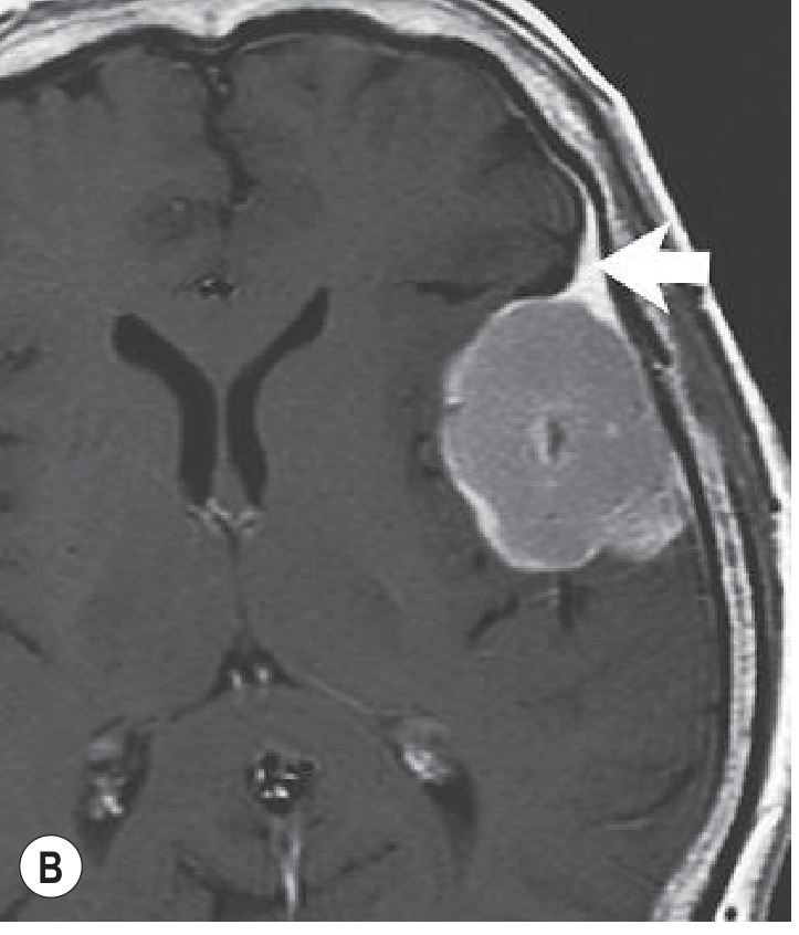
- Vivid and homogeneous enhancement on post-gadolinium sequences
- Exception: cystic or very densely calcified tumours produce foci of low signal within the mass
- 'Capping cysts' of CSF-equivalent signal intensity may be seen
- Vasogenic oedema is not infrequently associated; even small lesions can cause extensive oedema
- The presence of intra-axial oedema corresponds to an increased likelihood of recurrence
Fig. 55.27A - T2 axial MRI: A grey-matter isointense mass indenting the left cerebral convexity with displaced sulci and a small pial vessel (arrowhead) - classic extra-axial features:
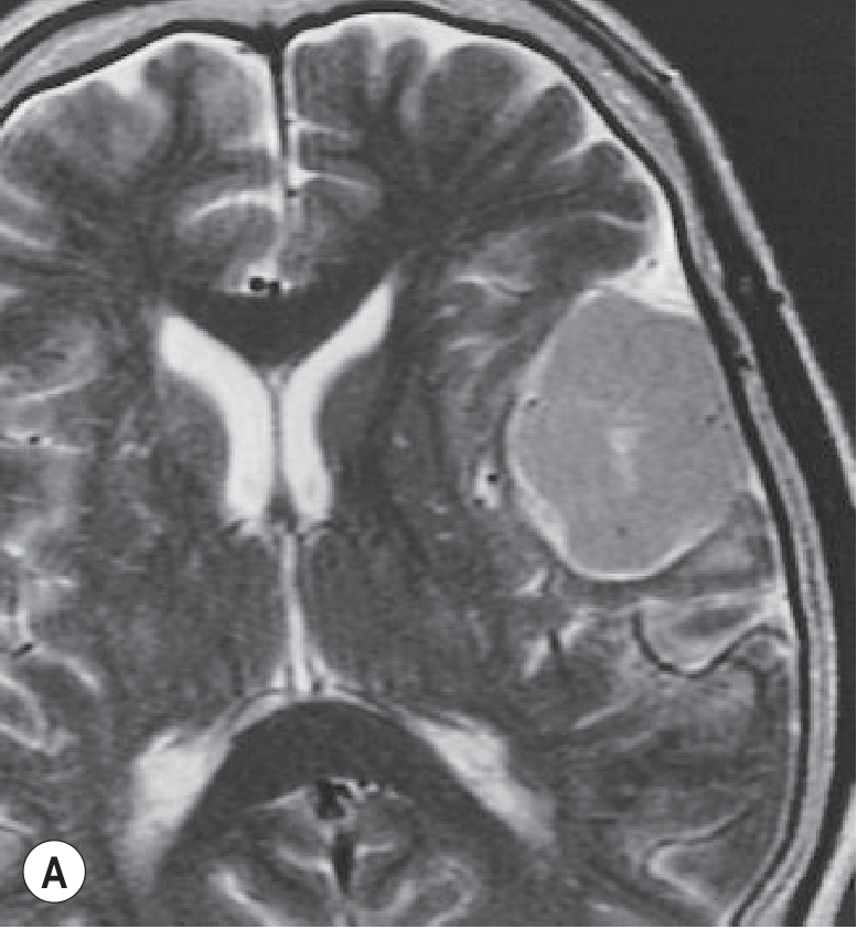
Fig. 55.27B - Post-gadolinium T1: Vivid homogeneous enhancement with the classic "dural tail" sign (arrow):
The "Dural Tail" Sign
- A linear, contrast-enhancing band extending from the tumour along the dura mater
- Once thought to be pathognomonic for meningioma
- Now known to also be seen with schwannoma and metastasis - not pathognomonic
Venous Sinus Involvement
Meningiomas abutting the superior sagittal or transverse sinuses can compress or invade these structures. Distinguishing compression from occlusion is critical for preoperative planning and can be assessed with MRA or CTA.
Advanced / Physiological MRI
MR Spectroscopy (MRS)
- May show an alanine peak - characteristic for meningioma
- Seen in less than 50% of cases
- No MRS data have been clinically correlated to patient outcomes
Diffusion-Weighted Imaging (DWI)
- Used to differentiate typical from atypical meningiomas, but results are conflicting
- Earlier reports suggested lower ADC values in non-typical meningiomas; more recent studies do not show a significant correlation
Perfusion-Weighted Imaging (PWI)
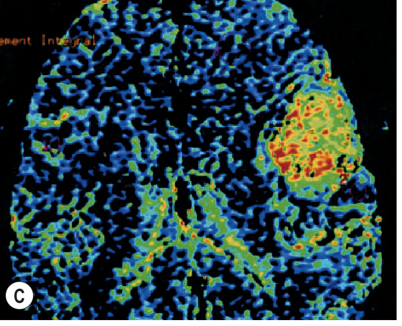
- Meningiomas usually have a markedly elevated rCBV - can differentiate from dural metastases, which tend to have lower rCBV
- CBV of peritumoural oedema is higher when surrounding malignant meningiomas
Fig. 55.27C - Perfusion-weighted MRI (colour rCBV map): Markedly elevated cerebral blood volume in the tumour (red-orange cluster) compared with normal cortex - confirming its highly vascular nature:
PET Imaging
- Meningiomas show high uptake of ⁶⁸Ga-DOTATOC (somatostatin receptor PET)
- Useful for:
- Differentiating meningioma from other dural-based tumours
- Distinguishing residual/recurrent tumour from postoperative enhancement
- Radiotherapy planning for skull base meningiomas
Angiography
- Now mostly performed for preoperative embolisation to minimise intraoperative blood loss
- Cardinal findings:
- Blood supply from meningeal vessels
- A dense, homogeneous, persistent tumour blush
- Parasitisation of cortical vessels (convexity tumours) or ophthalmic artery branches (subfrontal masses) is not rare
- Post-embolisation MRI: typical decrease in enhancement and reduced perfusion of the devascularised segment
Intraventricular Meningioma
- The commonest cause of a mass in the trigone of the lateral ventricle after the first decade of life
- CT and MRI appearances are similar to extraventricular meningiomas:
- Well-defined, globular lesion
- Usually hyperdense on CT
- May give similar signal to cerebral cortex on T1 and T2
- Marked contrast enhancement on both CT and MRI
Spinal Meningioma
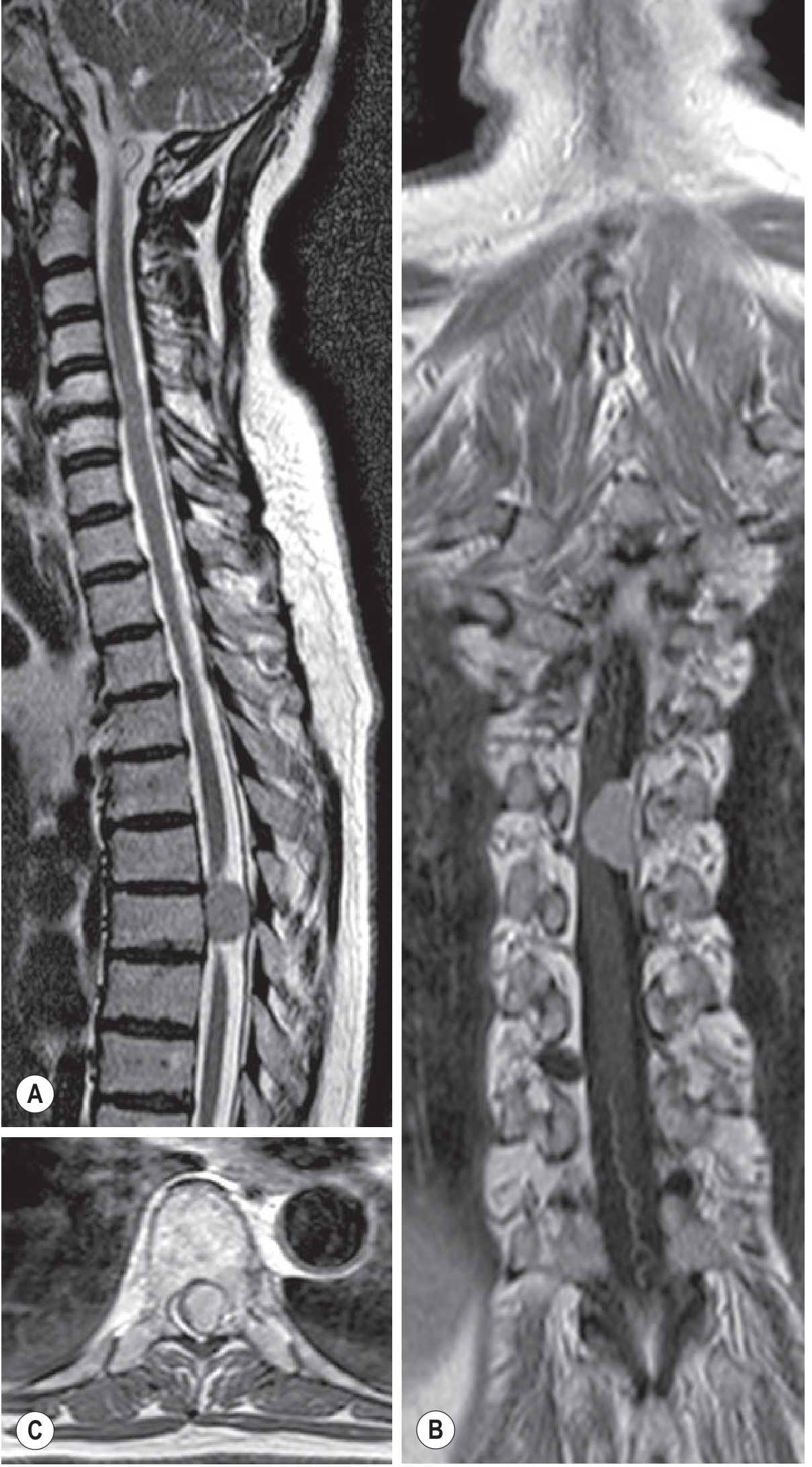
An intradural extramedullary tumour, commonly in the thoracic spine. MRI features (Fig. 49.19):
- Slightly hyperintense to spinal cord on T2
- Avid contrast enhancement on gadolinium-enhanced T1
- Broad dural attachment with a dural tail
- Can cause severe spinal canal narrowing and cord compression
Optic Nerve Sheath Meningioma
- Arises from the arachnoid sheath of the optic nerve (primary) or spreads from intracranial tumour (secondary)
- Most frequent in middle-aged women; bilateral = feature of NF2
- Imaging features:
- Generalised circumferential or fusiform enlargement of the optic nerve-sheath complex
- Calcification in 20-50% of cases on CT - relatively specific (not seen in optic nerve glioma)
- "Tram-track sign": peripheral enhancement of the tumour around the nerve on post-gadolinium fat-suppressed MRI (or hypoattenuation of the nerve on CT) - not specific; also seen in pseudotumour, optic neuritis, sarcoid, lymphoma, and metastatic disease
Differential Diagnosis
| Feature | Favours Meningioma | Favours Haemangiopericytoma |
|---|---|---|
| Shape | Spherical, broad dural base | Lobulated |
| Calcification | Present (up to 20%) | Absent |
| Hyperostosis | Common | Absent |
| Flow voids | Absent/rare | Multiple (highly vascular) |
- Infratentorial location: bone sclerosis favours meningioma; enlargement of the internal auditory meatus favours schwannoma
- Dural tail: also seen in schwannoma and metastasis
Grainger & Allison's Diagnostic Radiology, Chapter 55 (Extra-Axial Tumours) and Chapter 49 (Spinal Tumours) and Chapter 60 (Orbital Tumours)
This is a shared conversation. Sign in to Orris to start your own chat.