
Mark repeated questions Long notes in abdomen 1)rectus sheath 2)inguinal canal 3)stomach 4)spleen 5)liver 6)pancreas 7)duodenum 8)kidney 9)lesser sac 10)supra renal 11)rectum 12)anal canal 13)uterus 14)urinary bladder 15)prostate 16)ischio rectal fossa 17)portal vein and portocaval anastomosis 18)appendix Short notes 1)inguinal hernia 2)stomach bed 3)ligaments of liver and spleen 4)the bare area of liver 5)anterior and post relations of kidney 6)neck of pancreas 7)coeliac trunk 8)lesser omentum 9)epiploic foramen 10)hepatorenal pouch 11)porta hepatis 12)sigmoid mesocolon 13)the mesentry 14)pouch of doughlas 15)constrictions of ureter 16)positions of appendix 17)supports of uterus 18)superficial perineal pouch 19)deep perineal pouch 20)perineal body 21)urogenital diaphragm
anatomy abdomen repeated questions exam long notes short notes topics
Abdomen Anatomy - Repeated Questions Marked
LONG NOTES
| # | Topic | Status |
|---|---|---|
| 1 | Rectus sheath | |
| 2 | Inguinal canal | |
| 3 | Stomach | ⭐ Related to Short Note: Stomach bed |
| 4 | Spleen | ⭐ Related to Short Note: Ligaments of liver and spleen |
| 5 | Liver | ⭐ Related to Short Notes: Ligaments of liver, Bare area of liver, Porta hepatis, Hepatorenal pouch |
| 6 | Pancreas | ⭐ Related to Short Note: Neck of pancreas |
| 7 | Duodenum | |
| 8 | Kidney | ⭐ Related to Short Note: Anterior and posterior relations of kidney |
| 9 | Lesser sac | ⭐ Related to Short Notes: Epiploic foramen, Lesser omentum |
| 10 | Suprarenal | |
| 11 | Rectum | |
| 12 | Anal canal | |
| 13 | Uterus | ⭐ Related to Short Note: Supports of uterus |
| 14 | Urinary bladder | |
| 15 | Prostate | |
| 16 | Ischiorectal fossa | |
| 17 | Portal vein and portocaval anastomosis | |
| 18 | Appendix | ⭐ Related to Short Note: Positions of appendix |
SHORT NOTES
| # | Topic | Status |
|---|---|---|
| 1 | Inguinal hernia | ⭐ Related to Long Note: Inguinal canal |
| 2 | Stomach bed | ⭐ Related to Long Note: Stomach |
| 3 | Ligaments of liver and spleen | ⭐ Related to Long Notes: Liver, Spleen |
| 4 | Bare area of liver | ⭐ Related to Long Note: Liver |
| 5 | Anterior and posterior relations of kidney | ⭐ Related to Long Note: Kidney |
| 6 | Neck of pancreas | ⭐ Related to Long Note: Pancreas |
| 7 | Coeliac trunk | |
| 8 | Lesser omentum | ⭐ Related to Long Note: Lesser sac |
| 9 | Epiploic foramen | ⭐ Related to Long Note: Lesser sac |
| 10 | Hepatorenal pouch | ⭐ Related to Long Note: Liver |
| 11 | Porta hepatis | ⭐ Related to Long Note: Liver |
| 12 | Sigmoid mesocolon | |
| 13 | The mesentery | |
| 14 | Pouch of Douglas | ⭐ Related to Long Notes: Uterus / Rectum |
| 15 | Constrictions of ureter | |
| 16 | Positions of appendix | ⭐ Related to Long Note: Appendix |
| 17 | Supports of uterus | ⭐ Related to Long Note: Uterus |
| 18 | Superficial perineal pouch | ⭐ Related: Perineal body, Urogenital diaphragm |
| 19 | Deep perineal pouch | ⭐ Related: Perineal body, Urogenital diaphragm |
| 20 | Perineal body | ⭐ Related to Short Notes: Superficial & deep perineal pouch |
| 21 | Urogenital diaphragm | ⭐ Related to Short Notes: Superficial & deep perineal pouch |
Summary: High-Priority Repeated Clusters
| Cluster | Long Note | Short Note(s) |
|---|---|---|
| Liver | Liver (LN-5) | Ligaments of liver, Bare area of liver, Hepatorenal pouch, Porta hepatis |
| Kidney | Kidney (LN-8) | Anterior & posterior relations of kidney |
| Pancreas | Pancreas (LN-6) | Neck of pancreas |
| Stomach | Stomach (LN-3) | Stomach bed |
| Spleen | Spleen (LN-4) | Ligaments of liver and spleen |
| Appendix | Appendix (LN-18) | Positions of appendix |
| Uterus | Uterus (LN-13) | Supports of uterus |
| Inguinal region | Inguinal canal (LN-2) | Inguinal hernia |
| Lesser sac | Lesser sac (LN-9) | Lesser omentum, Epiploic foramen |
| Perineum | Ischiorectal fossa (LN-16) | Superficial perineal pouch, Deep perineal pouch, Perineal body, Urogenital diaphragm |
Study tip: Topics repeated across both sections are the examiner's favorites. For each Long Note, the paired Short Note is usually a sub-component - so mastering the Long Note covers the Short Note too. Focus hardest on Liver (4 short note overlaps) and Perineum (4 short note overlaps).
5 marks exam oriented short note on porta hepatis
Porta Hepatis
Definition and Location
Structures Passing Through
Entering the liver:
| Structure | Position |
|---|---|
| Portal vein | Posterior (behind both artery and duct) |
| Hepatic artery proper (right + left branches) | Left/medial |
| Lymphatic vessels | Along the structures |
| Hepatic nerve plexus (autonomic) | Along the vessels |
Leaving the liver:
| Structure | Position |
|---|---|
| Right and left hepatic ducts | Anterior/right lateral |
Memory aid: "PAD" from back to front - Portal vein (posterior), Artery (left lateral), Duct (right lateral/anterior)
Arrangement within the Hepatoduodenal Ligament
- Bile duct - right lateral and anterior
- Hepatic artery proper - left lateral
- Portal vein - posterior to both
Relations
- Anteriorly: Hepatoduodenal ligament (free edge of lesser omentum)
- Posteriorly: Epiploic foramen (foramen of Winslow) - the gateway to the lesser sac
- Below: First part of duodenum
- The structures at the porta hepatis are accessible by opening the hepatoduodenal ligament - this is the basis of the Pringle's manoeuvre (clamping of the hepatoduodenal ligament to control liver bleeding)
Lymphatic Drainage
- Hepatic nodes along the porta hepatis
- Then to the foraminal node at the epiploic foramen
- Then to superior pancreatic nodes and coeliac nodes
Clinical Importance
| Condition | Relevance |
|---|---|
| Cholangiocarcinoma (Klatskin tumor) | Tumor at the bifurcation of hepatic ducts at the porta hepatis - causes obstructive jaundice |
| Liver transplantation | Porta hepatis is the key surgical anastomotic site |
| Hepatic resection | "Lowering the hilar plate" gives access to porta hepatis for safe dissection |
| Pringle's manoeuvre | Clamping hepatoduodenal ligament at porta hepatis controls hepatic inflow during surgery |
| Biliary atresia (Kasai procedure) | Portoenterostomy is performed at the porta hepatis |
Exam tip: The most frequently tested fact is the arrangement of the three structures (duct - right, artery - left, vein - posterior). Always mention the hepatoduodenal ligament as the peritoneal envelope and the epiploic foramen as the key posterior relation.
5 Marks Short note on sigmoid mesocolon
Sigmoid Mesocolon
Definition
Shape and Root (Attachment)
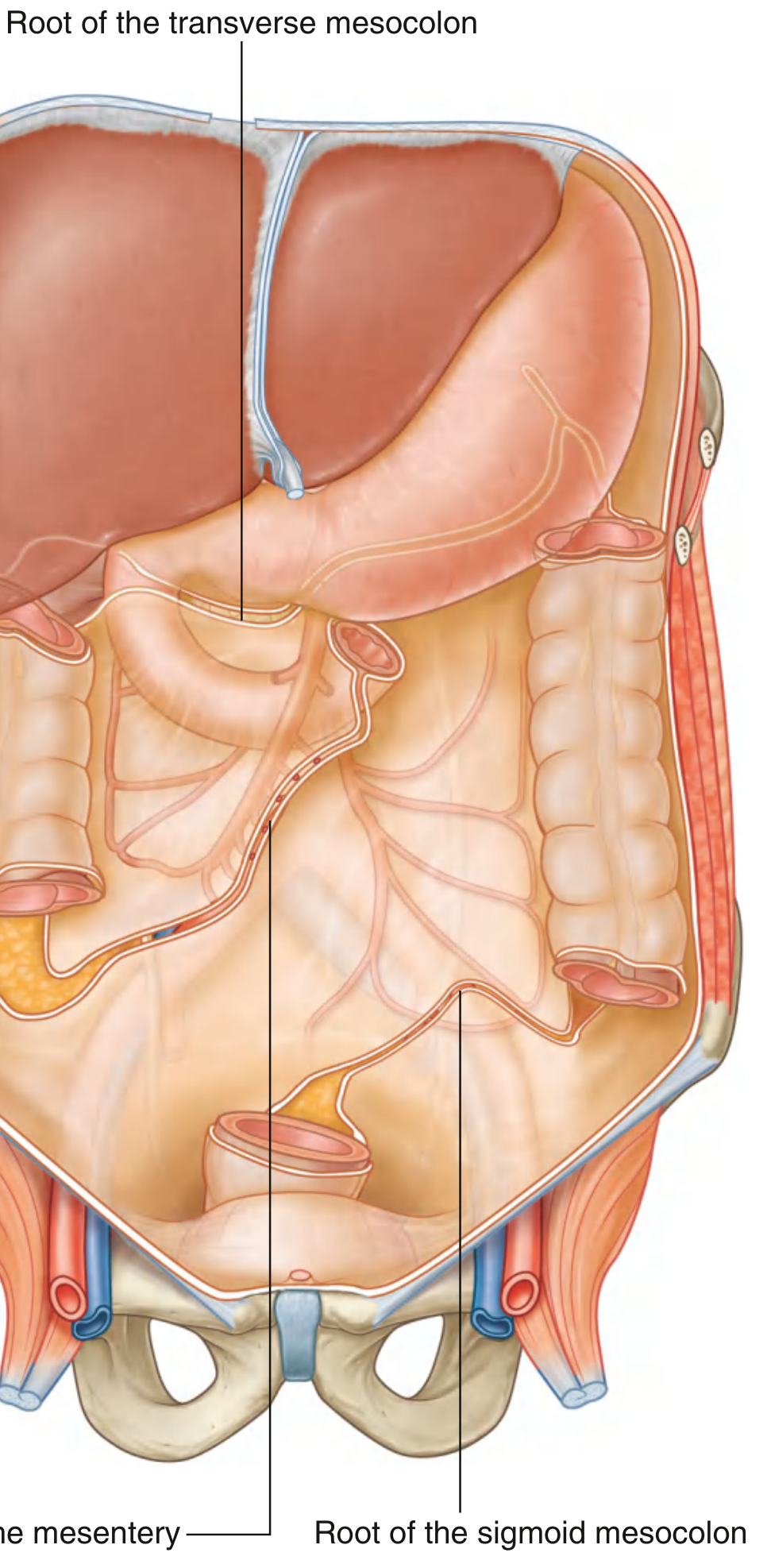
| Limb | Course |
|---|---|
| Left limb | Runs along the medial border of the left psoas major muscle (descending from above) |
| Right limb | Descends into the pelvis, ending at the level of vertebra S II (sacral vertebra 2) |
| Apex | Located near the bifurcation of the left common iliac artery into internal and external iliac branches |
Intersigmoid Recess
- At the apex of the inverted V of the root, there is a peritoneal pocket called the intersigmoid recess (intersigmoid fossa)
- It is a blind-ending peritoneal recess
- The left ureter lies immediately behind (retroperitoneal) at this point, running under the attachment of the mesocolon at the apex
- Clinically important: the left ureter is at risk during sigmoid colon mobilization - this mesenteric fold serves as the surgical landmark for the underlying left ureter
Contents
| Structure | Detail |
|---|---|
| Sigmoid arteries | 2-4 branches from the inferior mesenteric artery (IMA), descending to the left within the mesocolon |
| Superior rectal artery | Terminal branch of IMA, also runs in the lower part |
| Sigmoid veins | Drain into the inferior mesenteric vein |
| Lymphatics | Sigmoid lymph nodes drain along these vessels to paraortic nodes |
| Autonomic nerves | Sympathetic and parasympathetic fibers to sigmoid colon |
| Fat tissue | Fills in around the vascular structures |
Relations of Note
- The left ureter crosses under the apex of the inverted V (retroperitoneal)
- The left common iliac vessels lie at the apex
- The inferior mesenteric artery (IMA) runs close to the mesocolon and must be identified during surgical mobilization
Clinical Importance
| Condition | Relevance |
|---|---|
| Volvulus of sigmoid colon | The long mobile mesentery allows the sigmoid colon to twist on itself (most common site of colonic volvulus) |
| Left ureter injury | During sigmoid resection, the intersigmoid fossa marks where the left ureter lies; failure to identify it leads to inadvertent ureteric injury |
| Internal hernia | Loops of small intestine or greater omentum can enter the intersigmoid recess, producing an internal hernia |
| Diverticular disease | The long, mobile sigmoid with its mesocolon is the most common site for colonic diverticula |
| Sigmoid colectomy | The mesocolon must be divided close to the bowel wall or at its root depending on whether the surgery is for benign or malignant disease (oncological resection requires ligation of IMA at origin) |
Exam tip: The two most tested facts are (1) the inverted V-shape of the root with the apex at the left common iliac bifurcation, and (2) the intersigmoid recess containing/overlying the left ureter - this is the classic surgical landmark. Always mention the risk of ureteric injury.